An unusual group of children born with a defect or absence of one or both ears is called congenital microtia. According to the monitoring data of my country’s Birth Defect Monitoring Center, the incidence of birth defects in my country has remained high in the past ten years. Among them, congenital microtia is second only to cleft lip and palate, and there is about 1 “small ear angel” in every 7,000 people.
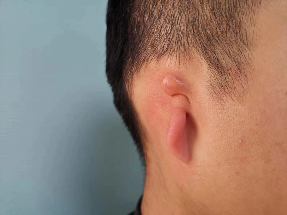
Congenital microtia is an important type of outer and middle ear deformity, which is a congenital structural birth defect. Manifested as born without ears or with partial auricle shape, peanut-shaped, sausage-shaped or leaf-shaped. Microtia is often associated with external auditory canal atresia or stenosis, middle ear deformity, and related organ deformities. The ear is as important as other organs as the window to listen to sound. Once the deformity occurs, it will have adverse effects on the child’s appearance, hearing function and mental health.
The treatment of congenital microtia mainly includes two aspects, one is the reconstruction of the auricle, and the other is the reconstruction of the auditory function. Reconstruction of the auricle is divided into two stages. The first stage is to reconstruct the external auricle, and the second stage is to erect the reconstructed “living ear” auricle. The repair surgery for atresia of the ear canal is usually performed in the middle of the two-stage operation of the auricle, creating a “live” ear for the child and opening the ear canal to rebuild the hearing function.
Auricular reconstruction challenges complexities within complexities
Microtia is not life-threatening, but it can affect the appearance and even the psychological development of children. With the development and innovation of modern medical plastic technology, auricular reconstruction has become the most effective method for the treatment of microtia, and it is one of the most challenging surgeries in plastic surgery.
The ear is the most complex body surface organ with the three-dimensional structure of the human body. Its shape and structure are very complex and have subtle concave-convex structures. During the operation, these fine structures must be reconstructed as accurately as possible, so as to achieve Symmetrical and beautiful effect. The selection of the auricle stent is the most important part of reconstruction. Generally, the self-rib cartilage is carved into the stent structure of the auricle. According to the growth and development law of children in my country, generally after the child is 7 years old, the chest circumference reaches 55-60 cm, and the ribs are 55-60 cm long. Surgery is performed when the cartilage has grown to a certain stage. At this time, the costal cartilage has good texture and elasticity, and can withstand a certain pressure. The affected ear can be individually sculpted and reshaped according to the shape and size of the healthy ear to make the shape more realistic. Reconstruction of the auricle not only requires mastering the carving techniques and skills of auricular stents, but also involves the application of skin grafting, skin flaps, and fascial flaps.
Auricular reconstruction requires a high level of professionalism and is very challenging for doctors. Currently, there are very few hospitals in the province that can successfully perform this type of surgery.
Atresia of the ear canal can be difficult to repair
The anatomy of the ear includes the pinna, external auditory canal, tympanic membrane, ossicles, and inner ear. The plastic repair of patients with atresia of the ear canal greatly tests the professional skills and surgical experience of experts. During the operation, the patient’s original ear canal needs to be ground out, the bony atresia plate is opened, and the mobility and continuity of the ossicular chain are examined to see if the ossicular chain is intact. When the sound reaches the ossicular chain, it continues to transmit inward. If the ossicular chain is fixed, poorly connected or interrupted, the sound transmission efficiency will be greatly reduced; if the ossicular chain is mobile, it is more conducive to surgery. After the sound is transmitted to the inner ear, the sound transmission efficiency is improved, which is more conducive to the improvement of hearing. The tympanic membrane of patients with osseous atresia of the ear canal needs to be taken from their own temporalis fascia and skin as supplementary materials to make a new tympanic membrane to form a complete sound conduction pathway.
Deformed ossicular chain is often fixed to the blocking plate. During the process of opening the bony blocking plate and using a drill to grind the blocking plate, noise may enter the inner ear and cause sensorineural hearing loss. , this injury should be minimized during the operation. Care should be taken to protect the facial nerve in the bone during otoplasty. The facial nerve innervates the facial muscles, which can make people show happy, sad and sad expressions. Avoid damage to the facial nerve during surgery to cause facial paralysis, so as not to affect the child’s future quality of life and social interaction.
The road is long and the road is coming. As one of the earliest units in China to carry out total auricle reconstruction, the Department of Otology of Shandong Otolaryngology Hospital has always been at the forefront and has accumulated rich experience in the treatment of ear deformities. Although a long way to go, but still handed in a satisfactory answer sheet.