3 treated babies born
Writing | Yan Xiaoliu
Source | “Medical Community” Public Account
Maternal-fetal medicine is another milestone.
Up to now, the first 3 fetuses with myelomeningocele (MMC) treated with intrauterine stem cells have been born successfully. One of them is one year old.
Relevant treatment occurs in the second trimester. Through an incision in the uterus, the surgical team places a biological patch containing special stem cells on the exposed spinal cord of the fetus to repair the surrounding protective tissue and reduce or prevent disabling complications.
“I’ve been working on this day for nearly 25 years.” Diana Farmer, the world’s first female maternal-fetal surgeon, led the study. She said so.
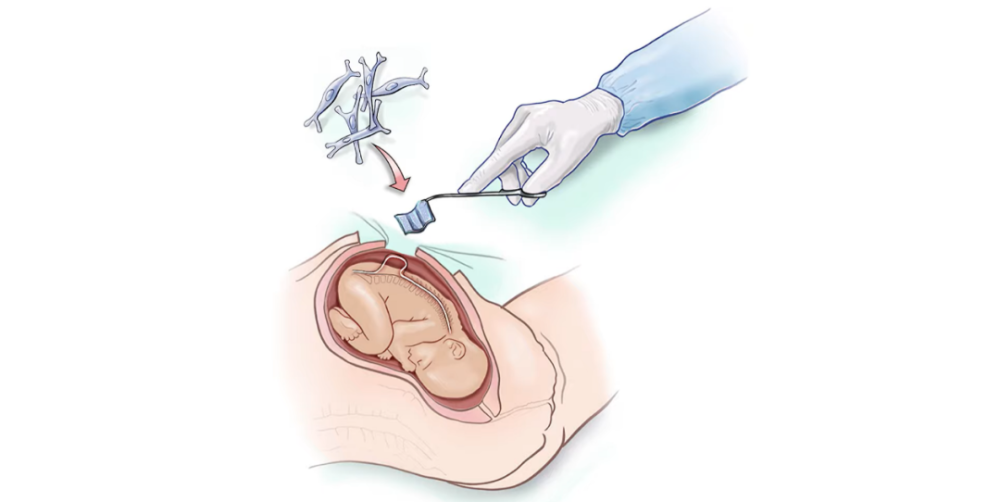
Image caption: Diagram of intrauterine stem cell therapy. /UC Davis
Find the “Path to Healing”
At 25 weeks and 5 days pregnant, Emily from Texas, USA, had “the most important surgery” of her life.
The subject of surgery is the fetus in the womb.
“My husband and I never thought that our baby would have developmental problems.” Emily recalled that her early pregnancy went well. However, the second-trimester ultrasound showed that the fetus had MMC.
This is a serious form of spina bifida, a birth defect. The specific manifestation is that part of the spinal cord and nerves are not covered by bone or skin, but are exposed. Many children suffer from cognitive, mobility, urinary and intestinal dysfunction and other complications, and may also cause brain damage due to hydrocephalus.
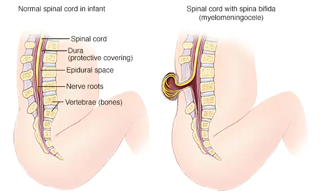
Image caption: On the left is the normally developing spinal cord and spine. On the right is a child with MMC. /Mayo Clinic
According to “Mayo Clinic” information, surgical treatment of MMC mainly includes prenatal and postnatal corrective surgery, which aims to put the exposed spinal cord of the fetus back in place, and then use patches, etc. Close the wound.
Early research confirms that longer spinal cord exposure is associated with greater damage and more and more severe complications. Therefore, prenatal surgery is expected to be carried out at around 26 weeks of gestation, either through laparotomy or under the fetoscope.
In the early 2000s, Diana Farmer led the clinical trials for the Myelomeningocele Management Study (MOMS). She found that prenatal corrective surgery was indeed effective in reducing neurological damage caused by MMC. But more than half of the patients were still unable to walk independently after treatment.
As principal investigator at the University of California, Davis, Diana Farmer hopes to find new ways to not only achieve local repair, but also to reverse the damage.
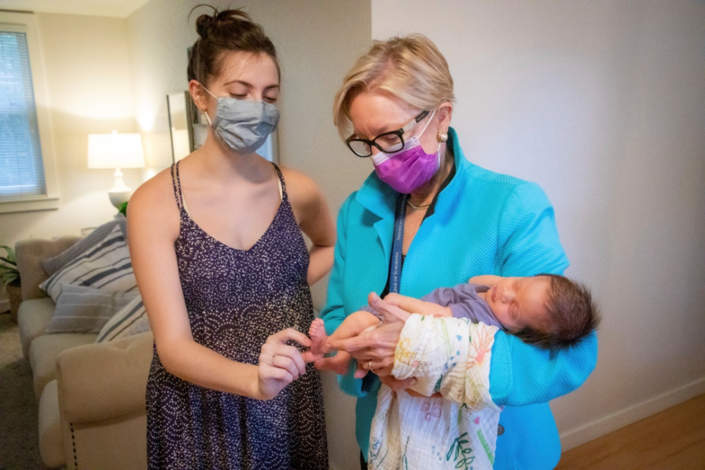
Image caption: Diana Farmer holds her baby after treatment and birth. /UC Davis
Animal experimentation is dawning
More than a decade ago, Diana Farmer and her colleague, Aijun Wang, a professor of biomedical engineering, established the Surgical Bioengineering Laboratory dedicated to stem cell and bioengineering research.
In its view, stem cells, as a class of cells with proliferation and differentiation potential, have good application prospects in regeneration and repair of damaged tissues. “The combination of cell therapy and surgery should promote tissue regeneration and help repair permanent damage.” Wang Aijun explained.
In 2015, the research team began to demonstrate the feasibility of cell therapy in animal experiments.
“We spent a lot of time deciding which cells to use and what animal models to build. We tried induced pluripotent stem cells, nanofiber scaffolds, tried various solutions protocol,” said Christopher Pivetti, director of the Surgical Bioengineering Laboratory.
In the end, the research team chose mesenchymal stem cells. This is an adult stem cell derived from the mesoderm in early embryonic development, with self-renewal, multi-directional differentiation potential, and through the bodyScale-up expansion still maintains its biological properties.
“We call them the ‘caregivers’ of the body because they produce healing factors that are ideal for repairing damaged tissues such as spina bifida. In contrast to embryonic stem cells, mesenchymal stem cells are used have fewer complications,” said Christopher Pivardi.
The team selected an approved dural patch as a biomaterial scaffold on which to place human placenta-derived mesenchymal stem cells. Then, in collaboration with the UC Davis School of Veterinary Medicine, through prenatal surgery, the patch was attached to the lamb’s congenital spina bifida. After the lamb was born, it was able to walk with no apparent disability.
Image caption: Dural patch. /UC Davis
Later, the team performed postpartum stem cell treatment for spina bifida on two bulldogs. The 4-month postoperative review showed that the dogs were able to walk, run and play independently.
The team believes that the related technology has achieved remarkable results in animals. “Not only is it safe, but it can also effectively improve walking ability. The nerve cytokines secreted by stem cells have successfully rescued the motor function of the animal model.”
On March 2, 2021, the University of California, Davis, announced the launch of the world’s first “Clinical Trial of Cell Therapy for Intrauterine Repair of Myelomeningocele” with FDA approval.
It’s called the CuRe Trial.
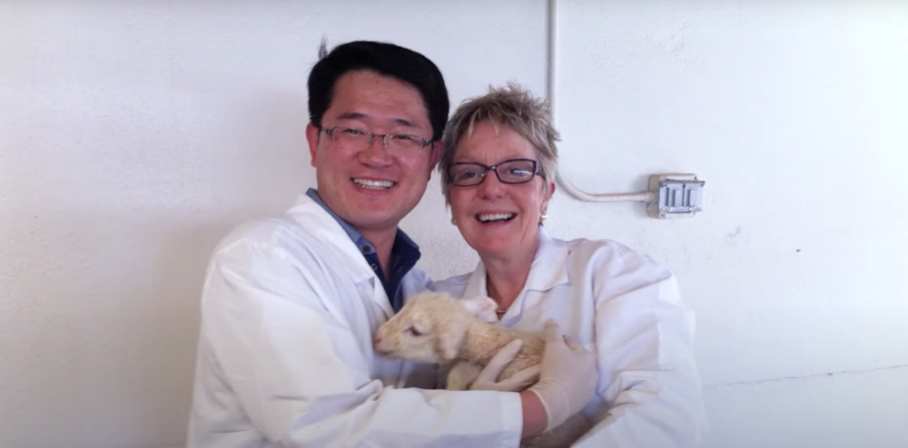
Image caption: Diana Farmer and Wang Aijun hug a healed lamb. /UC Davis
World’s first human trial successful
On the day she received her fetal MMC diagnosis, Emily heard about the “cure trial” for the first time.
“It’s a lifesaver we can’t refuse,” she said.
Fetal surgery is scheduled for July 12, 2021. More than 40 people participated in the process from the preparation of preliminary materials to the execution of the operation.
Before surgery, Wang Aijun and others extracted and produced clinical-grade mesenchymal stem cells from Emily’s placental tissue, and used them to make stem cell patches.
The Surgical Bioengineering Laboratory, Priya Kumar, leads the team to create the cell patch. “It takes 4 days. From removal of cells, to seeding on scaffolds, to delivery, every minute counts.”
On the day of the surgery, Emily was placed on her back in a hospital bed under general anesthesia.
The fetal surgeon makes a small incision in the uterus and carefully moves the baby’s back to the incision to fully expose the spine. Next, the cell patch is placed directly on the exposed spinal cord. Then, the incision is closed and the tissue is allowed to regenerate.
“The implantation went very well. Mom and fetus are great!” said Diana Farmer, the world’s first surgery to treat MMC with intrauterine stem cells.
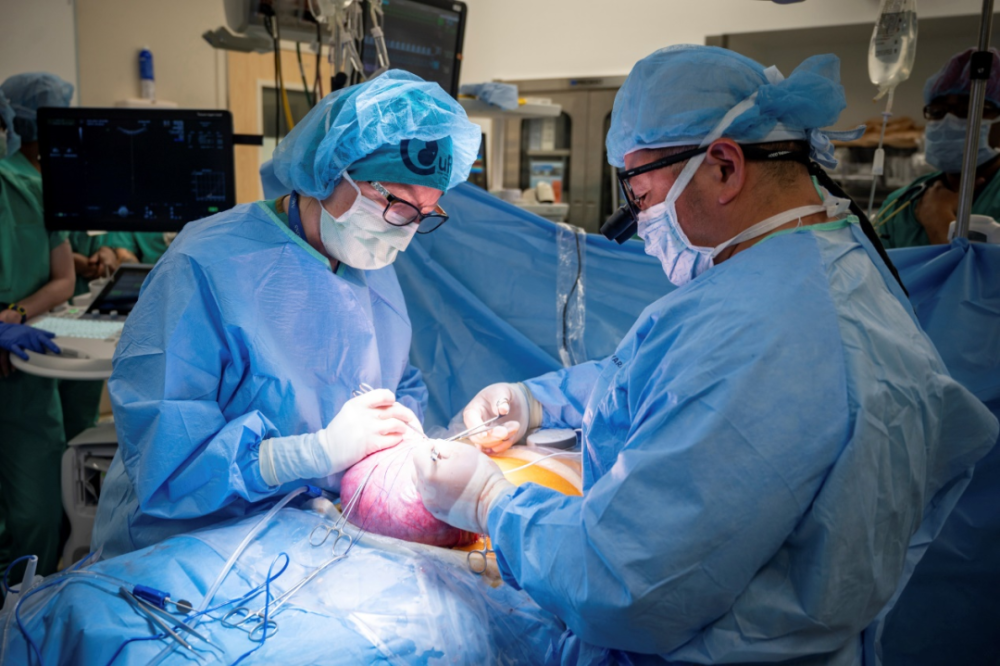
Image caption: During surgery. /UC Davis
At 35 weeks and 5 days of pregnancy, baby Robbie was born by caesarean section, weighing about 2.4 kg and measuring 48.26 cm.
“She was carried to me. I saw her toes wiggling,” Emily said.
“I vividly remember shouting out: Oh my God, she’s wriggling her toes! Did you see that!” said Diana Farmer, who has a degree of spina bifida. Very serious. Without intrauterine treatment, her legs may have been disabled at birth.
On September 20, 2022, Ruby celebrated her 1st birthday. Follow-up showed that she was in good physical condition.
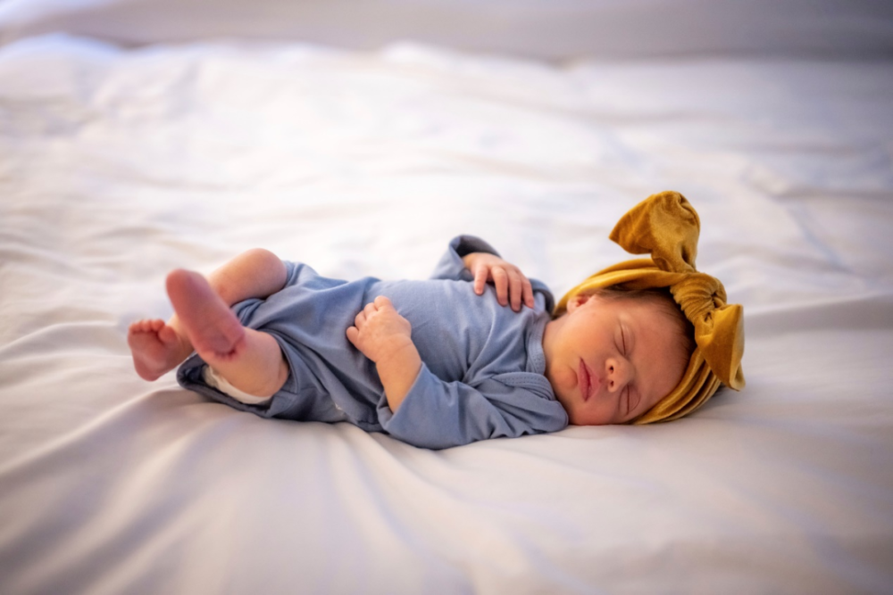
Image caption: Newborn Robbie. /UC Davis
The Cure Trial plans to enroll 35 MMC fetuses, the team said. The control group was MMC infants aged 0-4 months, and some underwent postpartum traditional surgical repair.
ResearchStudy follow-up will extend to 30 months of age. At that time, the babies will undergo key tests, including walking and toileting assessments, to fully assess the safety and effectiveness of the treatment.
Generally, children at this age have fully mastered major movements such as walking, running, jumping, standing, squatting, sitting, touching, and crawling.
Meanwhile, the research team will monitor their growth and development until age 6.
In recent years, the potential of stem cells to treat birth defects has gradually become known. According to some studies, placenta-derived mesenchymal stem cells can be made into “biological patches” to “patch” the underdeveloped heart. Other studies hope to use it to repair underdeveloped lung tissue.
“The research seems to be going well, but we are cautious and don’t jump to conclusions.” Diana Farmer said that every success story is peeling an onion, and there are bound to be bad ones . “Fortunately, we never gave up.”
“We are honored to be a part of making history,” she said.
References:
[1]World’s first stem cell treatment for spina bifida delivered during fetal surgery.UC Davis Health
[2]Stem cell patch surgery to mend spina bifida in the womb.BBC
[3]The CuRe Trial: Cellular Therapy for In Utero Repair of Myelomeningocele. Study Pages
[4]First stem cell clinical trial for spina bifida treatment announced.UC Davis Health
Source: Medicine
Proofreading: Zang Hengjia
Editor in charge: Tian Dongliang
* The medical community strives for the accuracy and reliability of its published content when it is reviewed and approved, but does not regard the timeliness of the published content, and the accuracy and completeness of the cited materials (if any), etc. Make any promises and guarantees, and do not assume any responsibility for the outdated content, possible inaccuracy or incompleteness of the cited information. Relevant parties are requested to check separately when adopting or using it as a basis for decision-making.