
Case data
A 42-year-old man presented to the emergency department complaining of nausea, vomiting, and abdominal pain for 1 week. On presentation, his condition worsened with partial loss of consciousness and signs of dehydration. The patient was initially diagnosed with acute renal insufficiency, and her condition improved after being hospitalized and receiving intravenous fluids. Three days later, the patient developed acute abdominal pain and was treated conservatively with broad-spectrum antibiotics and intravenous fluids, but the patient developed sepsis within a few hours. Laparotomy was performed, and gastric perforation was found during the operation. The operation was successful, and the drainage tube was retained in the upper abdomen. The patient’s condition was relieved, and when the abdominal drainage tube was removed 4 days later, a large worm was found stuck at the distal end of the drainage tube (Fig. 1), which was confirmed as yes based on its shape, color, length and shape (round; tapering to the end) A giant kidney worm (Nephrodontia). In addition, the patient was microscopically found to have three co-infections (P.
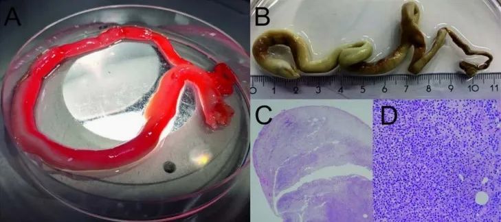
Figure 1 A, Freshly released worms before fixation in formalin B, worms after formalin fixation C, HE-stained histological cross-section showing the outline of the worm (enlarged 25x) D, HE stained histological cross-section showing extensive neutrophil infiltration and destruction of the internal structure of the parasite (200x magnification)
Analyze the discussion
Nephrodontia is a large parasitic nematode, commonly known as Meganephrosis, which is mainly parasitic on more than 20 kinds of animals such as dogs, minks, wolves, and Rattus norvegicus of the kidneys and intra-abdominal cavity, occasionally infecting humans, with only about 40 reported cases of human infection and the majority in Asian. Animals can become infected by ingesting oligochaete annelids (intermediate hosts, such as Hirudidae, etc.) containing the larvae of the second-stage nephron. Human infection is generally caused by eating raw or semi-raw frog or fish (transferred host) containing the third stage larvae of the worm, or by eating raw water or lettuce > Infection on an oligochaete host. This disease can be prevented by cooking food and not drinking raw water.
The adult worms usually exist in the kidney, and the worms excrete eggs in the urine. The clinical manifestations include fever, low back pain, hematuria, pyuria, etc. >Misdiagnosed as kidney tumors, urinary tract stones, and urinary tract infections. Diagnosis requires the detection of worms or eggs in the urine. The efficacy of medical therapy is unknown, and surgical resection is the preferred treatment in most cases.
A ectopic infection occurs when adult worms are found outside the kidney of the end host, as was the case in this patient. Ultrasound images of its kidneys showed no signs of destruction or infection, and molecular identification of the worms was unsuccessful, likely due to DNA degeneration due to formalin fixation and extensive leukocyte infiltration in the worms (Figure 1C and D). This suggests that the worms died long before they were removed from the abdominal cavity. Since gastric biopsy does not reveal other causes of gastric perforation (eg, Helicobacter pylori infection, malignancy, gastric ulcer, or gastroscopic ischemia), adult-induced gastric perforation may be from one side of the abdomen of live worms penetrate the stomach wall (which is subsequently killed by the resulting inflammatory response), or die adult worms in the abdominal cavity near the stomach (which subsequently causes an inflammatory response that thins the stomach wall and causes perforation). This is the first case of human intra-abdominal infection with Nephron nematode, a patient presenting with gastric perforation. This rare disease is easily misdiagnosed clinically, and attention should be paid to the recognition and differential diagnosis.
References:
1. Boerekamps A, Janmaat VT, Schrama YC, et al. First reported case of an ectopic renal giant worm (Dioctophyme renale) infection in the abdominal cavity. J Travel Med. 2022 Jul 14 ;29(4):taac015.
2. Carra Perera S, Silveira Mascarenhas C, Brum Cleff M, Müller G, Silva Rappeti JC. Dioctophimosis: a parasitic zoonosis of public health importance. Adv Exp Med Biol 2021; 1306:129 –42.
3. Lei Bo, Pang Yaqin, Kong Baoqing, Wang Fengyong. The first report of human nematode infection in Guangxi [J]. Journal of Youjiang Medical College for Nationalities, 2004(04):544.< /p>
4. Xiao Qing, Liu Xiaoxia, Zhao Xuelan, Zhao Hong, Wang Jinling. A case of nephrosis [J]. Chinese Journal of Nephrology, 2003(06):25.