Have you ever noticed in your daily life that you or others have small holes in your ears, the corners of your eyes, or around your nose, but you clearly remember that you have never been hurt or pierced by needles. What’s the matter?
Forget it, but sometimes I can’t help but squeeze my hands when I see it, so what are these holes? Is it really safe to squeeze? Is there any way to fix and fill these holes?
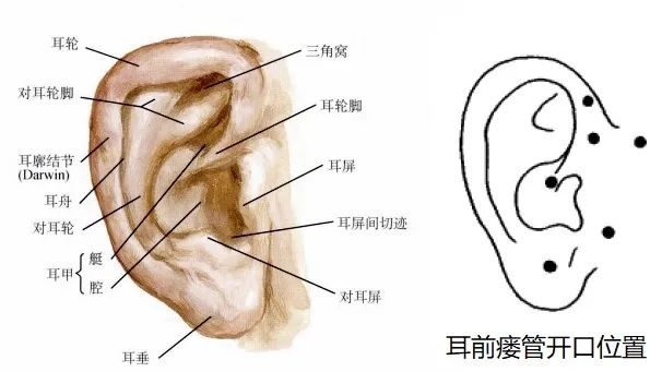
Small holes around the ears
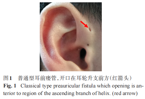
The small hole around the ear may be a preauricular fistula (congenital preauricular sinus tract), commonly known as the ear canal, and it is said that the hole in the ear represents wealth and honor Blessed to be smart, but it’s just a congenital malformation of the outer ear.
The specific pathogenesis is unknown, which may be related to abnormal embryonic development and the failure of the ear to close properly. Most of the disease is unilateral, and the incidence is generally higher in women than in men.
Preauricular fistulas can be divided into simple, secretory and infective according to clinical manifestations. Simple type usually has no symptoms, generally does not need treatment, keep it clean every day, don’t squeeze it at will, and don’t put earrings and earrings on it because it is a natural “ear hole”.
Secretory type refers to the accumulation of sebum and sebaceous secretions in the fistula, resulting in frequent white bean curd-like secretions and a peculiar smell on the fistula (hole) , can cause local skin itching, local bulge.
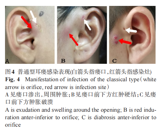
Infectious type (82.58%) showed local redness, swelling, pain, and pus discharge from the fistula; in severe cases, the surrounding soft tissue swelled, forming >Abscess ruptures or granulation, repeated infection persists for a long time, and scar is formed at the site of repeated infection.
The only effective way to prevent the recurrence of the preauricular fistula is complete surgical excision of the fistula tissue. The timing of surgery is generally selected after the acute infection is controlled, or it can be performed after the acute infection. Direct surgical resection.
Acute infections require systemic antibiotics. After the abscess is formed, it needs to be incised and drained in time, and the dressing should be changed regularly until the infection is controlled and scarring is formed. For the secretions produced, can be gently wiped with alcohol cotton pads, etc., do not squeeze with hands or poke with unclean toothpicks.
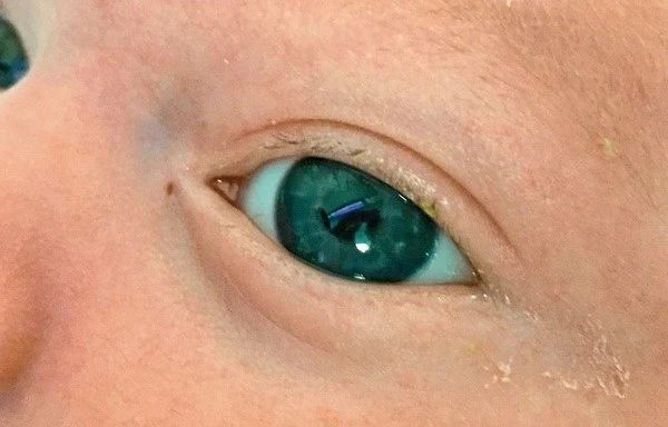
A hole next to the corner of the eye
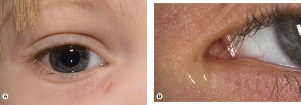
The skin of the lacrimal sac at the corner of the eye has a pinpoint-sized hole from which the tear fluid escapes. This condition is dacryocystorial fistula, which can be divided into congenital and acquired sex.
Congenital dacryocystorial fistula is a rare congenital anomaly of lacrimal duct development, the etiology of which is unknown, mostly sporadic, and occasionally familial. There are two modes of inheritance: autosomal dominant and recessive.
Acquired dacryocystorial fistula is mainly formed after the rupture of lacrimal sac inflammation, and there is inflammatory granulation tissue proliferation around the fistula.
Congenital dacryocystorial fistulas have small openings with neat margins and no inflammatory granulation tissue. It is mostly unilateral, and the fistula opening is usually located on the lateral side of the nose, below the medial canthal ligament, and it has also been reported to be located at the lateral canthus. Fistulas often overflow with clear fluid or mucus. In some children, the tearing symptoms disappear when the fistula is blocked by secretions.
If there is only one small hole and no other symptoms, it is not necessary to deal with it, and regular cleaning can be done. For symptomatic patients, fistula resection or combined dacryocystorhinostomy can be used after acute inflammation subsides.
Hole in nose
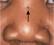
The small holes in the nose may be nasal dermoid sinuses. Nasodermoid sinuses and nasodermatoid cysts arise from congenital fusion abnormalities at the root of the nose.
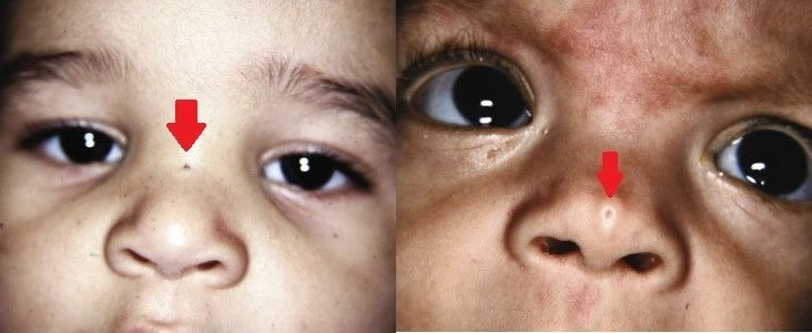
Nasal dermoid cysts are epithelial-lined sinuses that extend from the base or tip of the nose through the septum and into the anterior cranial fossa. Usually an incompressible mass on the dorsum of the nose, with a dimple or pit somewhere along the midline of the dorsal surface of the nose.
Nasal dermoid cysts with intracranial extension increase the risk of CNS infection and should be surgical excision. The conventional wisdom is that a craniotomy, usually a staged operation, is required.
It is now believed that for lesions involving the skull base or slightly involving the intracranial, endoscopic-assisted minimally invasive surgery to reach the skull base + direct rhinotomy or open rhinoplasty ( That is, from the tip of the nose to the back of the nose) treatment.
Today is another day full of knowledge and dry goods. Come and see if you or your friends have these small holes~ If you want to know any secrets about your body, welcome Leave a commentOw~
[References]
[1] Zhou Ping, Chen Jinhui, Huang Ting, et al. Research progress of congenital preauricular fistula [J]. Journal of Clinical Otolaryngology Head and Neck Surgery, 2019(5):4.
[2] Zhu Yaying, Li Chenlong, Shi Yuxuan, et al. Progress in diagnosis and treatment of congenital preauricular fistula [J]. Chinese Journal of Ophthalmology and Otolaryngology, 2019, 019(001):11-15 .
[3] Wang Dong, Luo Wugen. Observation and surgical treatment of congenital preauricular fistula [J]. Chinese Journal of Otology, 2020, 18(4):4.< /p>
[4] Wei Mingzhuang, Luo Qining, Huang Jiayun, et al. Clinical features and surgical treatment of infected preauricular fistulas[J]. Chinese Journal of Otology, 2021, 19(1):5.< /p>
[5] Wei Mingzhuang, Luo Qining, Huang Jiayun, et al. Clinical features and surgical treatment of infected preauricular fistulas[J]. Chinese Journal of Otology, 2021, 19(1):5.< /p>
[6]Glenn C Isaacson, MD, FAAP. Congenital Nasal Anomalies. UpToDate