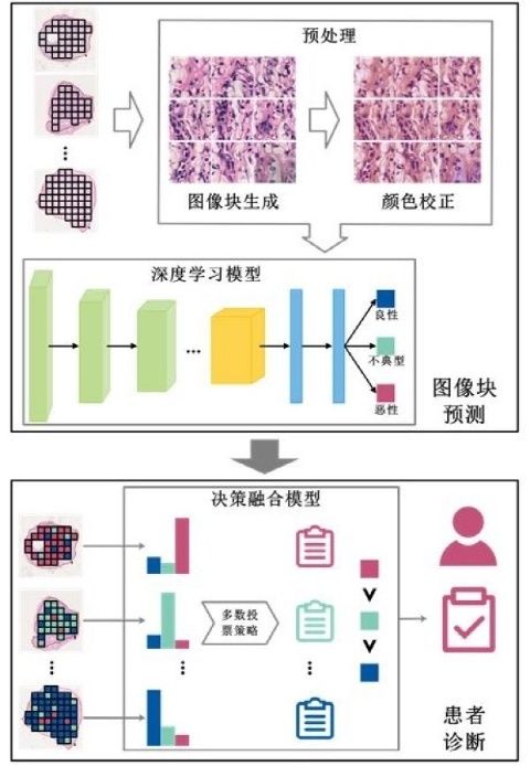
Skin cancer is one of the most common malignant tumors. Among them, melanoma has the strongest invasive ability and the highest degree of malignancy, is prone to lymph node and hematogenous metastasis, has a high mortality rate, and seriously threatens human health. Melanoma is a malignant melanocytic lesion, and melanocytic lesions include atypical and benign lesions. Clinically, the treatment and prognosis of different types of melanocytic lesions are different. Patients with malignant melanocytic lesions (melanoma) need to undergo surgical resection, combined with adjuvant therapy such as radiotherapy and chemotherapy, interferon therapy and immunotherapy; patients with atypical melanocytic lesions only need surgical resection of the lesions without adjuvant such as radiotherapy and chemotherapy Treatment, but close follow-up observation is required; patients with benign melanocytic lesions only need to remove the lesions. Therefore, early and accurate diagnosis of melanocytic lesions is of great significance for the formulation of surgical plans and the improvement of patient prognosis. Currently, the clinical diagnosis of melanocytic lesions is mainly through histopathological analysis, which relies on the experience of the pathologist, is highly subjective, time-consuming, and has a high rate of inconsistency (average inconsistency rate of 45.5%). Benefiting from the maturity of whole slide scanning technology, Computer Aided Diagnosis (CAD) based on pathological whole slide image (WSI) can provide a solution to the above problems. In recent years, artificial intelligence (AI) technology has made many breakthroughs in the field of CAD based on pathological WSI, but the existing pathological diagnosis of melanocytic lesions based on AI has not yet achieved the identification of atypical melanocytic lesions, and atypical melanocytic lesions The surgical plan of the patient is different from that of patients with benign and malignant melanocytic lesions, but the histological patterns and biological characteristics of atypical melanocytic lesions partially overlap with those of benign and malignant melanocytic lesions, and are easily confused with benign and malignant melanocytic lesions (Figure 1). In response to the above problems, the Suzhou Institute of Biomedical Engineering and Technology of the Chinese Academy of Sciences and the Department of Pathology of the Ninth People’s Hospital Affiliated to Shanghai Jiaotong University School of Medicine have proposed a new method for fully automatic and intelligent pathological diagnosis of melanocytic lesions (Figure 2). The study included 711 patients with melanocytic lesions from 3 centers—374 with benign lesions, 119 with atypical lesions, and 218 with malignant lesions. The scientific research team uses the deep learning method to construct an image block prediction module, outputs the probability of melanocyte lesion type, and realizes the objective and quantitative digital interpretation of the local information of the pathological tissue slices; the decision fusion strategy is used to aggregate the prediction results of all image blocks of each melanocytic lesion patient. Thereby, a patient diagnosis module is constructed. The results show that the accuracy of the method is 96.3% and 93.0% on the internal and external test sets, respectively, which is significantly higher than that of three clinical pathologists (two senior pathologists with more than 10 years of experience in pathological diagnosis and one freshman). The accuracy rate of independent diagnosis of junior pathologists who have completed 3 years of standardized training); in addition, with the assistance of this method, the diagnostic accuracy of pathologists has been improved, especially the diagnostic accuracy of junior pathologists has improved by nearly 40.0%. This study explores and verifies the clinical application potential of AI technology in assisting pathologists to improve the diagnosis accuracy of melanocytic lesions, and provides a new way to improve the current situation of severe shortage of pathologists in my country. At present, the research team is using related technologies to carry out intelligent and precise diagnosis research in ophthalmology and neuropathology, and is committed to building a set of integrated solutions for multidisciplinary and multi-disease digital pathology intelligent diagnosis. Related research results were published in the Journal of Dermatological Treatment. The research work was supported by the National Natural Science Foundation of China and the Shanghai Municipal Health Commission.

Source: Voice of Chinese Academy of Sciences