[Source: Huaihua News Network]
Recently, the intervention team of the Department of Cardiovascular Medicine of the First Affiliated Hospital of Hunan Medical College successfully carried out several cases of intravascular ultrasound imaging (IVUS) after the IVUS machine was installed. Under the guidance of percutaneous coronary intervention (PCI) surgery, the hospital opened a new journey of precise interventional diagnosis and treatment of coronary heart disease, improved the scientificity and accuracy of interventional treatment strategies, made coronary interventional treatment more objective and accurate, and benefited the majority of patients.

Preoperative angiography
The 72-year-old Uncle Guo was independently completed by the Department of Cardiovascular Medicine of the hospital. The first patient undergoing PCI under the guidance of IVUS, Uncle Guo showed severe coronary stenosis in recent angiography, including 50% stenosis in the proximal segment of the left anterior descending artery and 90% stenosis in the middle segment. There are indications for PCI. In order to reduce the risk of surgery and improve the prognosis of the patient, it is recommended that Uncle Guo complete the operation under the guidance of IVUS, and inform him of his condition, necessity and risks of the operation. Uncle Guo and his family agree.
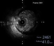
remote foothold
 < /p>
< /p>
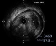
The narrowest part of the lesion
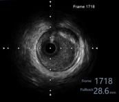
Proximal landing site
Passed IVUS inspection It was found that Mr. Guo had diffuse stenosis in the proximal and middle segments of the anterior descending artery, the plaque load at the most stenotic point was 85%, and the vascular lumen area was only 2.4 m2. (Choose the stent size, length and the best place for stent implantation), successfully implanted a 3.5×29mm drug-eluting stent for Uncle Guo. After repeated IVUS optimization of stent inner diameter and wall adhesion, IVUS was performed again before the end of the operation. Inspection: The stent adhered well and expanded well, without dissection, thrombus, or plaque prolapse, and the minimum lumen area of the anterior descending stent was 7.82 m2. All data met the requirements after stent implantation, and the operation was successfully completed.
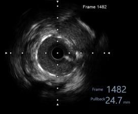
Minimum lumen area after stent implantation
According to Director Yu, with endovascular Ultrasound (IVUS) is the “eye” under the guidance of cardiovascular physicians to complete complex and high-risk PCI operations. It not only provides surgeons with accurate information on the nature and stenosis of intravascular lesions, but also provides accurate information on vascular lesions. The judgment of length and blood vessel diameter is more accurate, and it also provides an accurate objective basis for judging the need for stent implantation, stent adherence, and surgical risk assessment. IVUS has greatly improved the level of interventional diagnosis and treatment of coronary heart disease, and greatly guaranteed patient safety. . (Gu Dan and Jiang Taoqian)
Disclaimer: The copyright of this article belongs to the original author. If the source is wrong or your legal rights are violated, you can contact us by email, and we will processed in a timely manner. Email address: [email protected]