Otitis media is a common problem, but there may be other “hidden things” behind it. Ms. Chen has been plagued by otitis media for more than two years, and it is very disturbing. It was only recently that the root cause of the disease was found to be a tumor at the base of the skull next to the nasopharynx. Fortunately, the otorhinolaryngology medical team of Ningbo Huamei Hospital of the University of Chinese Academy of Sciences (Ningbo Second Hospital) took a different approach and completely removed the large tumor buried in the paranasal skull base, which relieved the hidden danger of the patient.
Hearing stuffiness, hearing loss, otitis media
This tumor is very cunning and hidden in the base of the skull
Ms. Right ear fullness and hearing loss. “When the problem occurred, the right ear seemed to be covered with a piece of cloth. It was unbearably uncomfortable!” Since then, the otolaryngology department has become the place where Ms. Chen reports every now and then. She was diagnosed with “secretory otitis media” at the local hospital, and she tried taking medicine and puncturing the tympanic membrane. Every time she felt more comfortable after treatment, and it didn’t take long for her ear stuffiness and discomfort to come back!
In this way, Ms. Chen suffered for more than two years, and finally found a “right nasopharyngeal space occupying” through nasopharyngeal CT in a superior hospital.
Is it a tumor? This made Ms. Chen’s family pale in shock and came to the outpatient service of Wang Kai, the head of the Otolaryngology Department of the Huamei Hospital of the National Science and Technology University. After a comprehensive examination of nasopharyngeal CT, MRI plain scan + enhancement, etc., the diagnosis was “paranasal skull base tumor”.
“This tumor is very cunning, close to the base of the skull, and hidden deep!” Wang Kai introduced that the human ear and the nasopharynx are connected by the Eustachian tube, and the secretions in the ear flow into the Eustachian tube. Nasopharyngeal discharge. The patient’s tumor compresses the right Eustachian tube, which prevents smooth discharge of secretions and causes repeated otitis media. Different from oropharyngeal tumors in lower position, huge tumors are located in the paranasal skull base and high in position. This makes the nasal endoscopy, only the right nasopharyngeal round pillow can be found to bulge more than the left, allowing this tumor to slip away cunningly again and again!
The tumor is surrounded by blood vessels and nerves
In the face of difficult cases, the expert team innovated the surgical route
The special location of the tumor not only delayed the diagnosis, but also provided treatment. brings difficulty. There are major blood vessels such as the internal carotid artery and external carotid artery, as well as a series of important nerve tissues such as the facial nerve, glossopharyngeal nerve, and hypoglossal nerve around the tumor. If the tumor is allowed to continue to grow, Ms. Chen will not only face repeated episodes of otitis media, but the tumor will also destroy the bone at the base of the skull upwards, and invade the nerves and face downwards and outwards.
“But the tumor grows high and hides deeply. The traditional surgical approach is either poor exposure of the tumor or too much trauma.” Focusing on this difficult case, Wang Kai led the team to conduct a careful preoperative evaluate. If you follow the surgical approach for common oropharyngeal tumors, whether it is from the mouth or sawing the mandible, the surgical path is long and the trauma is very large; if the incision is made from the right earlobe to the back of the neck, the tumor will be removed from the back to the front, although the trauma is small However, the intraoperative visual field is poor and the risk is too great.
With rich surgical experience in ear neurosurgery and ear-lateral skull base surgery, Wang Kai boldly proposed a new surgical approach—infratemporal fossa A-type approach. “Simply put, it is to make an incision behind the right ear to the back of the earlobe, then continue to the neck, and pass through the internal carotid artery and external carotid artery from the side to find the tumor.” The surgical path proposed by Wang Kai is generally applied to the temporal bone. For tumors, tumors invading the jugular foramen and other lesions, no one has tried to remove paranasal skull base tumors in eastern Zhejiang.
Preoperative multidisciplinary careful design plan
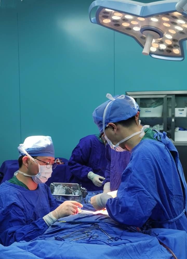
3-hour operation, “dig out” the large tumor deep in the skull base
Before the operation, the otorhinolaryngology department invited The radiology department, ICU, etc. conducted multidisciplinary discussions, and formulated a detailed and thorough surgical plan and related treatment measures. After being fully prepared, Wang Kai’s team performed the operation on the patient for more than three hours. During the operation, the surrounding nerve trunks and major blood vessels were perfectly exposed and isolated.
The blood supply around the tumor was abundant, and the otolaryngology team was walking on thin ice during the operation. In order to accurately touch the position of the tumor at the base of the skull and avoid massive hemorrhage due to damage to the blood vessel wall when surgical instruments are removed, Wang Kai used his fingers to probe into the gap between the internal carotid artery and the external carotid artery, and after clearing the tumor boundary, he used it under a microscope. The nerve stripper bluntly separated the tumor, and slowly “pulled out” the tumor with a diameter of about 3 cm, and successfully removed the time bomb buried deep in the patient’s skull base.
After surgery, Ms. Chen recovered well. Due to the meticulous protection of the surrounding tissues such as the skull base and nerves during the operation, the patient did not experience complications such as cerebrospinal fluid leakage, cranial nerve palsy in the posterior group, and facial paralysis. He recovered and was discharged from the hospital one week later.