Recently, the team of Professor Wang Ning, director of the Second Neurosurgery Ward of the First Affiliated Hospital of Harbin Medical University, successfully removed a rare case of a giant pituitary adenoma with multiple sites of invasion by using the combined endoscopic-microscope technique.
The 48-year-old Ms. Dong began to feel unwell 2 months ago. At first, she just thought that she had reached menopause and her body function was degraded, but then her body discomfort continued to worsen, and she was accompanied by decreased vision in her left eye. , performed an MRI of her head at a local hospital and found a huge tumor in her intracranial pituitary region. After many inquiries, he found Professor Wang Ning for treatment.
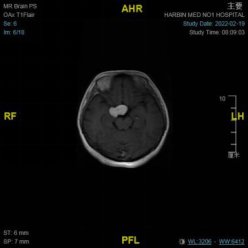
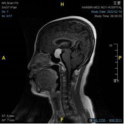
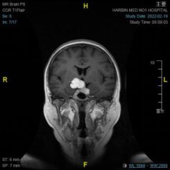
Professor Wang Ning understood the medical history and made a diagnosis of giant pituitary adenoma. Because the patient’s tumor is huge, 4 cm, and the upper part grows to the right, it is impossible to achieve complete resection through nasal endoscopic surgery;
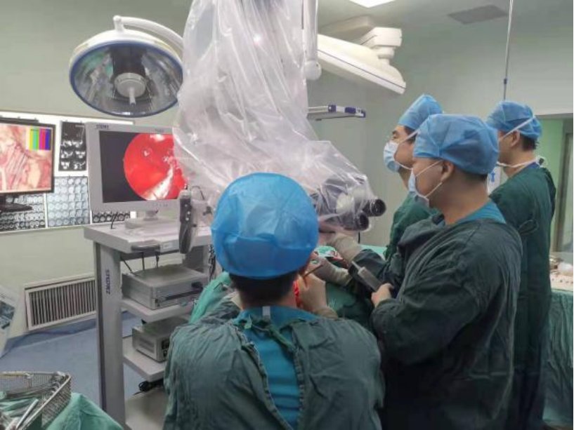
Professor Wang Ning’s team completed the preoperative examination for the patient. After systematic evaluation and consideration, they decided to adopt an individualized surgical plan. ——The domestic leading endoscope-microscope combined technique is Ms. Dong’s tumor resection. With the cooperation of the anesthesiology department and the operating room, Professor Wang Ning led the attending physician Shao Qi, and the residents Fang Yulong, Kong Fanyi, and Wang Hongfei to successfully remove the tumor for the patient.
Professor Wang Ning introduced that at present, many neurosurgical skull base tumor centers in China use transcranial or staged surgery for the resection of this type of tumor. Update, only a few skull base tumor centers in China have carried out the first-stage “double-lens combined surgery”. This combined operation is performed by two groups of doctors, who use a microscope to perform transcranial and neuroendoscope transnasal synchronously. The endoscopic transnasal tumor is removed, and the craniotomy part uses a minimally invasive concept to release the tumor from the tightly adhered important blood vessels and nerves. It is freed and pushed into the endoscopic field of view, which not only protects nerves and blood vessels, but also facilitates complete tumor resection.
Professor Wang Ning pointed out that the application of the dual-lens combined technology realizes the complementary advantages of the neuroendoscope and the microscope in the same operation, and makes full use of the depth, flexible angle and close observation of the neuroendoscope. The stereoscopic field of view under the microscope and the advantages of precise separation of blood vessels and nerves on the tumor interface under direct vision allow for safe tumor treatment, avoiding the technical deficiencies and risks that may be brought about by a single surgical method.
Shao Qi Chen Ye