All media reporter Tan Qionghui correspondent He Lei Du Juan
“Thanks to the doctors in the Department of Cardiology of Minda Hospital, you gave us new life!” Recently, Minda Hospital Affiliated to Hubei University for Nationalities He took the lead in independently carrying out OCT surgery with optical interference tomography in the state. After the patients recovered, Zhang Changjiang, the director of the Department of Cardiology, was “blocked” in the office to express his gratitude to them.
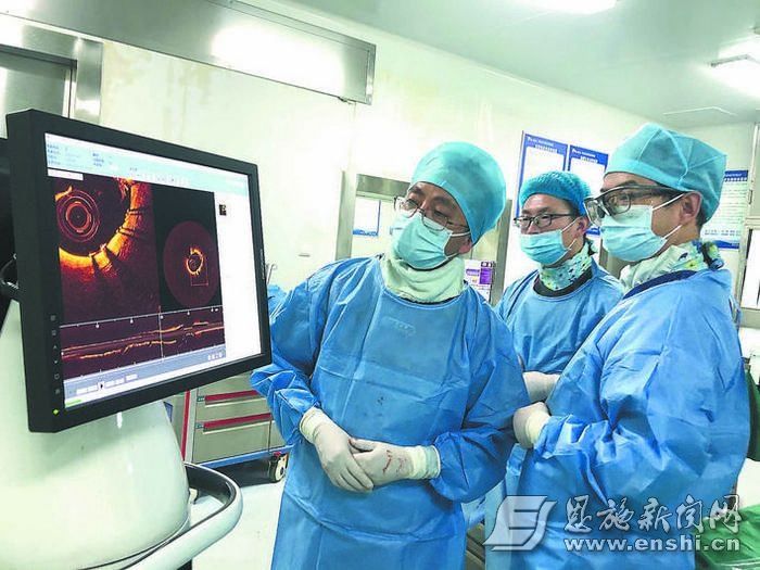
OCT in progress.
On New Year’s Eve, she suffered from episodic palpitation, chest tightness, and fatigue… The 58-year-old Ms. Yu’s “old problem” relapsed. She came to the Department of Cardiology of Minda Hospital for treatment. After inquiring about the medical history, the doctor diagnosed her as complex coronary vascular disease based on the examination results, requiring precise interventional examination and treatment.
Department of Cardiology Director Zhang Changjiang and Deputy Director Li Huabo organized the intervention center staff after careful discussion, and completed angiography through a guiding catheter. It was found that the patient had diffuse lesions from the opening to the middle of the anterior descending artery, and the blood vessels were combined with obvious calcification, and the most severe stenosis was 80%. %, the circumflex branch is also a diffuse lesion, the right coronary artery is irregular throughout, distal chronic occlusion, and long calcification lesions, which again increases the difficulty and risk of surgical operations, and increases the risk of postoperative thrombosis.
The team led by Zhang Changjiang immediately used optical coherence imaging catheter OCT-guided revascularization. The OCT catheter with high-resolution and rapid imaging is the “eye of the sky” for the interventional cardiologist, which ensures the expansion and good adherence of the stent, and reduces the risk of postoperative thrombotic events. After stent implantation, OCT examination showed that the stent expanded well throughout the whole process, and the operation of the branch stent was perfect.
Zhang Changjiang introduced that OCT imaging technology allows doctors to visually see everything in the patient’s blood vessels, just like a “real eye”, and can provide the safest and most effective treatment plan for the patient’s follow-up treatment. In the OCT examination, the three-layer structure of the intima, media and adventitia of Ms. Yu’s blood vessel can be clearly seen. The stent is tightly “attached” to the coronary artery, and the metal stent beam is clearly visible.
3 days after the operation, Ms. Yu recovered and was discharged from the hospital.
Coincidentally. Not long ago, Mr. Sun, a citizen, had a reexamination of vascular lesions and had repeated chest pains for many years.
Because of the complexity of vascular lesions, Zhang Changjiang repeatedly communicated and explained with his family members, and finally the family members agreed to do interventional examination and treatment. The angiography results showed that the left main trunk of the patient had no obvious stenosis, the anterior descending artery had severe stenosis of about 70%, the opening of the circumflex branch was about 80%, and the middle and distal right coronary artery was occluded. The interventional team used optical coherence imaging catheter (OCT)-guided revascularization again, implanted 3 stents, and successfully performed the operation.
In recent years, coronary angiography has developed from pure coronary angiography to a combination of coronary angiography with functional imaging and intraluminal imaging. At the end of last year, Minda Hospital independently carried out QFR (quantitative blood flow fraction) to evaluate the interventional guidance of coronary physiology function, combined with OCT application technology, marking that Minda Hospital’s coronary intervention technology has entered a new era of precise treatment, which will be a great opportunity for the Wuling area. More coronary heart disease patients bring good news.
What exactly is “Eye of the Sky”?
OCT, also known as optical interference tomography, is the imaging technology with the highest resolution at present, with a resolution of up to 10 microns (1 millimeter equals 1000 microns), commonly known as optical biopsy. The new “gold standard” for the diagnosis of coronary heart disease.
OCT test is to place an imaging catheter with an optical lens at the head end in the coronary artery, and through high-speed rotation and retraction, it can help us diagnose the structure and plaque inside the blood vessel in less than 3 seconds. The block nature is like putting the eye directly into the blood vessel to see, and the whole diagnosis process is safe and reliable.
The biggest advantage of OCT is that it has a high resolution of up to 10 microns, which can accurately identify plaque stability and detect plaque rupture.
Stable plaques have thicker fibrous caps, while unstable plaques, i.e. vulnerable plaques, are thin-capped fibrous atheromas rich in inflammatory cells, like “thin-skinned, large-stuffed rotten plaques.” Meat dumplings”. The characteristic of vulnerable plaque is that it is very easy to rupture. Once the plaque ruptures, it is easy to immediately form a thrombus to block the blood vessel, causing acute myocardial infarction, and even life-threatening and sudden death. OCT detection can detect vulnerable plaques in time and prevent the occurrence of acute and malignant cardiovascular events as soon as possible.
What is the role of OCT?
Although coronary angiography is a common and effective method for diagnosing coronary heart disease, it is only a two-dimensional plane image, which cannot allow us to understand what is happening in the blood vessels, and has certain limitations.
OCT greatly complements coronary angiography and can identify normal vessels, lipid plaques, fibrous plaques, calcified plaques, thrombi, dissections and intimal tears, as well as other intravascular Pathological images, such as vulnerable plaques with thin fibrous caps, macrophages, microvascular channels in the vessel wall, etc., are some vascular pathological changes that cannot or cannot be clearly seen in angiography and other examination tools.
Its functions can be briefly summarized into the following three aspects:
Pre-stent: reveal the morphology and properties of coronary plaques, distinguish calcification, fibrous and lipid plaques, Vulnerable plaque was found, red and white thrombus were identified, and stent size was accurately selected;
During stenting: immediate adherence of the stent was observed, and acute thrombosis, intimal tear and plaque prolapse were found;
p>
After stenting: follow-up to understand stent endothelial repair, intimal hyperplasia, late thrombosis and the progress of bioabsorbable stent absorption.