Reporter Li Xiwei Correspondent Zhu Guotao
A few days ago, the Cardiology Department of the Second People’s Hospital of Jining carried out new technology again and completed the first intravascular ultrasound examination. The application of this technology marks that hospital cardiac interventional therapy has entered the era of precise treatment, and also provides a stronger diagnosis and treatment guarantee for patients with coronary heart disease.
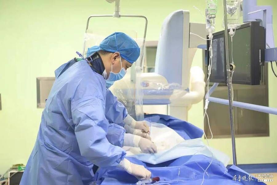
Patient Shao Moumou was admitted to the Cardiology Department of Jining Second People’s Hospital due to episodic chest pain and chest tightness for 20 years. Received coronary angiography + stenting. After coronary angiography, the patient’s left main trunk and anterior descending artery had severe stenosis. For precise treatment, the hospital’s Cardiology Interventional Team performed an intravascular ultrasound examination on the patient, and accurately measured the patient’s left main trunk. The minimum cross-sectional area of 11 square millimeters , the minimum cross-sectional area of the middle segment of the anterior descending branch is 1.9 square millimeters. Subsequently, the expert team placed a stent in the mid-section of the LAD with the most severe stenosis. The LM did not need to place a stent. The postoperative intravascular ultrasound clearly showed that the patient’s blood vessels were unobstructed and the stent adhered well.
The main cause of coronary heart disease is coronary atherosclerosis, which is a chronic disease formed by the accumulation of waste in blood vessels. Although traditional coronary angiography has always been regarded as the “gold standard” for evaluating coronary lesions, this method also shows many shortcomings in clinical application, such as only the lumen can be displayed, and the wall and plaque where the lesions are located cannot be displayed. It is very likely that doctors underestimate the degree of coronary artery disease, and some complex lesions cannot accurately judge the degree of stenosis.
Intravascular ultrasound (IVUS) detection technology is to send a hair-thin ultrasound probe into the target blood vessel, perform 360° scanning in the blood vessel cavity, and clearly display the cardiovascular structure and vascular structure through the display screen. disease. This technology can further examine the coronary lumen on the basis of angiography, and “see” the coronary artery lesions more carefully. It can not only quantitatively measure and analyze the diameter of blood vessels and the degree of stenosis, but also clarify the shape, nature and distribution of coronary lesions. As an important supplementary method for coronary angiography, IVUS largely makes up for the lack of visual analysis of the severity of lesions, improves the accuracy of lesion diagnosis, and avoids the need for empirical excessive stent implantation. PCI) strategy, stent selection and effect evaluation have important guiding significance.
In the next step, the Cardiology Department of Jining Second People’s Hospital will continue to practice the “patient-centered” service concept with practical actions, and continuously bring new concepts and new technologies at the forefront of the profession to the clinic. Continue to optimize and improve the medical technology level of the team to provide more high-quality and efficient medical services for more patients.