Qilu Evening News Qilu One Point reporter Yu Wei correspondent Li Qian Sinan
The eyeball with retinopathy caused by diabetes is filled with a dense proliferative membrane, which causes the patient to see things unclear or even blind. How to strip the culprit of this disease and help patients regain their light is a huge test for ophthalmologists.
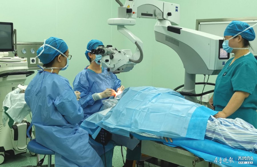
As an ophthalmology department of Jining First People’s Hospital with a history of 86 years, it has been committed to the forefront of industry technology, taking the lead in using the 25G+ cutting speed high-speed vitrectomy system as a conventional vitrectomy treatment plan, reducing the In addition to the complications of conventional vitrectomy, it has greatly improved the surgical experience of patients with diabetic retinopathy, retinal detachment, macular hole, epimacular membrane, etc., and opened a new era of precise vitrectomy, which is brought to more than 300 patients with fundus diseases every year. bright.
Feng Jie, director of the Department of Ophthalmology of Jining First People’s Hospital, introduced that the vitreous body is a filler in the eye, similar in shape to jelly, and filled into the eye to play a supporting role. Vitrectomy, as the name suggests, involves removing the vitreous so that surgical instruments can reach the diseased area on the retina.
The eye, as one of the most precise organs of human beings, operates in the eye, leaving only a few millimeters of room for ophthalmologists to perform, and the technical requirements of the doctor are extremely strict: the surgeon needs to perform an operation in the eye. On the soft and transparent retina with the thickness of only a white paper, the proliferative membrane with extremely tight adhesion was peeled off. The whole operation is like dancing on the tip of a needle, and if you are not careful, it will fall short. Vitrectomy, because of its difficult operation and high technical content, is a manifestation of the technical level and comprehensive strength of a hospital’s ophthalmology to a certain extent.
Feng Jie said that by introducing the cutting-edge 25G+ high-speed vitrectomy system in China, we use a vitrectomy knife to puncture into the vitreous body during the operation. Compared with the original, this new technology has greatly improved the cutting speed. , greatly shorten the operation time, and also significantly reduce the traction on the “negative film” of the eye – the retina, which can liquefy the vitreous faster and increase the suction flow rate. At the same time, the bevel incision head can be closer to the retina for refined operations, better deal with small proliferative tissues, greatly improve the efficiency of the operation, and relieve the tension of the patients during the operation. For patients, this high-speed vitrectomy technology has less incision and trauma, faster postoperative recovery, minimizes surgical complications, and improves the patient’s surgical experience.

Related Links:
Features of Ophthalmology Diagnosis and Treatment in Jining First People’s Hospital:
1. Vitrectomy for diabetic retinopathy, macular hole, epimacular membrane, complex retinal detachment and other complex fundus diseases.
2. Wide-field fundus examination instrument for screening of neonatal fundus lesions.
3. Cataract, pterygium and other surgeries will be discharged on the same day of surgery.
4. Capsular bag tension ring assisted in the treatment of complex lens insufficiency and dislocation, one-time implantation of intraocular lens.
5. Endoscopic nasal dacryocystorhinostomy and cannulation in the treatment of complex chronic dacryocystitis.
6. Treatment of congenital cataract, congenital ptosis and other congenital eye diseases.
7. Vision screening and correction for adolescents and children.
8. Diabetic fundus screening and multi-wavelength laser therapy.
9. Non-invasive optical correlation tomography angiography (OCTA) examination, fluorescein sodium retinal angiography and indocyanine green choroidal angiography double-like angiography examination to assist in the diagnosis and treatment of complex fundus lesions.
10. The intrascleral interlayer was fixed with no suture to suspend the intraocular lens.