The largest immune organ in humans is the spleen, which may have the largest surface area and is the mucous membrane. Some people think it is the skin, because there are also many important immune cells in the skin, such as Langerhans cells.
If the human body is compared to a kingdom, then the immune system is our “army” to defend our country, and this army has a clear division of labor and strict discipline.
The immune system is composed of immune organs, immune cells and immune molecules, among which the immune organs are divided into central immune organs (composed of bone marrow and thymus, immune cells differentiated according to their location) development sites), and peripheral immune organs (including spleen, lymph nodes, mucosa-associated lymphoid tissues, etc., where immune cells settle and generate immune responses) [1].
This time we are going to focus on a low-key and extremely important “immune organ”, that is the mucosa-associated lymphoid tissue.
What is mucosa-associated lymphoid tissue? The name is a little weird!
As you may know, lymphoid tissue is another name for immune tissue in our body, so what is “mucous membrane-related”?
Mucosal-associated lymphoid tissue actually refers to the lymphoid tissue distributed in the parts of the human body where there is a mucosa.
These mucosal sites include the respiratory tract, gastrointestinal tract, genitourinary tract, and the mucosa of some exocrine glands (salivary, lacrimal, and mammary glands) that perform local-specific immune functions main venue [2].
Choose to reside in the mucosa, what do these lymphoid tissues think?
To solve this problem, we first need to understand what a mucous membrane is.
The mucosal tissue is the place where our human body communicates with the external environment, which is equivalent to the outer wall of the human kingdom. For example, the inside of the eyelids, the inside of the mouth, the nasal cavity, and the gastrointestinal tract are covered with mucosal tissue.
They need to be in direct contact with external antigens, the so-called “aggressors” (such as food, bacteria, viruses, etc.) every day, and are the most threatened parts of the human body.
Over 95% of our infections are caused by “aggressors” invading or invading the mucous membranes [2].
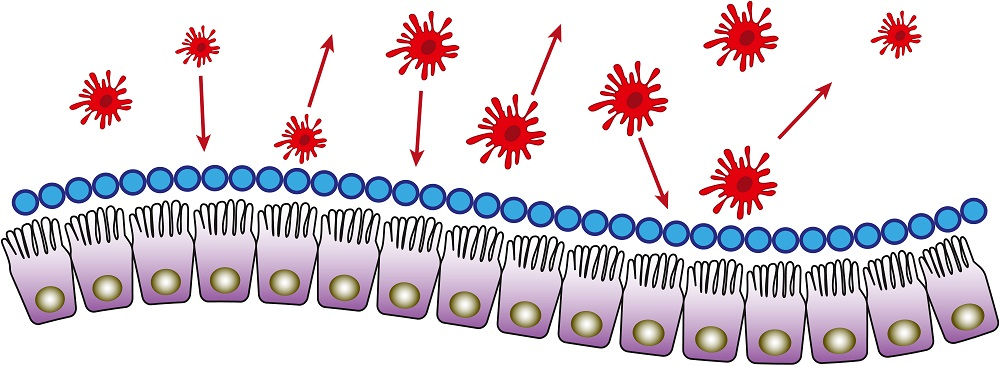
In order to prevent such an important part from being captured, the human body has deployed a large number of heavy troops to guard various parts of the city wall. We have given them a unified organization for this part of the army, which is called mucosa-associated lymphoid tissue (i.e. mucosal immune system).
Mucosal-associated lymphoid tissue forms a tight defense system, establishing the first immune barrier for the human body in the battle against the invasion of pathogenic microorganisms.
They come into play whenever a suspected “aggressor” invades, distinguishing whether these “aggressors” are harmless or harmful to decide whether to “let go” (medically It is called immune tolerance) or “blocked” (called immune response in medicine), once harmful microorganisms are found, they will prepare themselves for combat, and they will immediately report to the superior immune organization.
Mucosal immune tolerance is a complex concept, not simply “letting go”, but involving the participation of intestinal Treg cells, microbial flora (polysaccharides or TGF factors) , immune regulatory signals, etc.
Mucosal-Associated Lymphoid Tissue, who makes up this army?
Mucosal-associated lymphoid tissue is actually a general term for a type of lymphoid tissue, which refers to those lymphoid tissues distributed in the parts of the human body with mucosa, including the gastrointestinal tract, upper and lower respiratory tract, genitourinary ducts, exocrine glands (ie, lacrimal ducts, salivary secretory ducts), which occupy most of the human lymphoid tissue and play a very important role in the anti-infective immune response [2].
According to the different locations of these lymphoid tissues, they are named four categories, namely intestinal mucosa-associated lymphoid tissue (GALT), bronchial mucosa-associated lymphoid tissue (BALT), Conjunctival-associated lymphoid tissue (CALT) and urogenital mucosa-associated lymphoid tissue (UALT).
Mucosal-Associated Lymphoid Tissue, what does this army do?
The mucosal-associated lymphoid tissue is the “largest” of the immune system in terms of numbers, with more lymphocytes than the rest of the body combined.
60% of human lymphocyte T cells are located in mucosal tissues[2].
The unique location, composition and function of the mucosa-associated lymphoid tissue can protect the human body from pathogens (bacteria, viruses, etc.) On the other hand, it can make the human body develop immune tolerance to common food antigens and normal microorganisms, that is, let those harmless “aggressors” go (called immune tolerance in medicine). by).
How is the army of iron made?
Then, everyone must be very curious. Mucosal-associated lymphoid tissues are in front-line positions every day. They have to face a large number of external antigens that are mixed with mermaids and fishes. How do they achieve such an orderly and efficient manner? What about unmistakable identification and defense?
To understand an army, we first need to understand their team composition and corresponding role-playing.
Therefore, we firstLet’s take a look at the team composition of mucosa-associated lymphoid tissue.
For example – intestinal mucosa-associated lymphoid tissue (GALT)
We take the digestive tract as an example.The mucosa of the digestive tract is the place where nutrients are absorbed, digested and exchanged. 200 times the surface of the skin[2].
The mucosa-associated tissue (GALT) of the gut is composed of PP nodes (also known as intestinal aggregates), mesenteric lymph nodes, and various diffuse immune cells and their products, which cooperate with each other , and work together to complete the guardianship of this huge field.
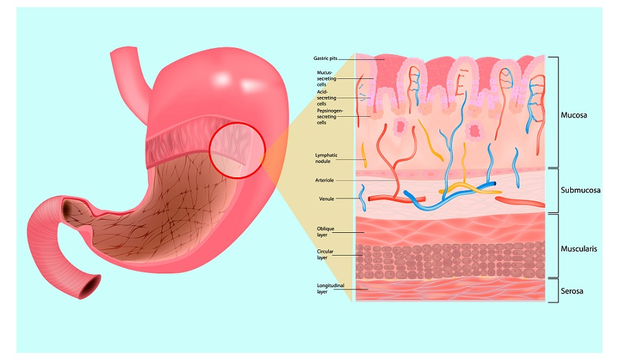
PP nodes – lymphocyte ‘bunkers’ on the mucosa of the small intestine
PP nodes, a group of lymphocytes in the mucosa of the small intestine, are equivalent to “bunkers”. There is only a layer of intestinal epithelial cells between them and the intestinal lumen.
The PP node is divided into two regions, the lymphoid follicle and the interfollicular region, usually guarded by B cells and T cells, respectively.
When harmful microorganisms invade,Microfold cells, commonly known as M cells (Microfold cells, a special type of intestinal epithelial cells, are the zones that antigens can pass through and can transfer antigens to Swallow it in the stomach and transport it to the “bunker” for identification) is responsible for delivering the message.
After the M cells send information, the B cells residing in the lymphoid follicles will proliferate and differentiate into plasma cells in a short period of time, and rapidly produce WMD secreted immunoglobulin (SIgA), which prevents microorganisms from adhering and multiplying in the mucosal epithelial layer, thereby preventing them from entering the epithelial layer.
At this time, T cells mainly located in the interfollicular zone are rapidly transformed into CD4+T cells and CD8+T cells (mainly CD4+T cells here) , personally into the battle.
 Source: Zhanku Hailuo
Mesenteric lymph nodes – “transit points” between mucosal and peripheral immune systems
Mesenteric lymph nodes are the lymph nodes located in the mesentery. They are equivalent to post stations. They transmit the news of the enemy’s invasion to the superior troops in time, so that they can get timely reinforcements.
Mesenteric lymph nodes are the transit points between the mucosal and peripheral immune systems [2]. What is the so-called transfer station?
Because the mesenteric lymph node is located in the backbone of the hub, it is connected with the PP node on the one hand, which is convenient for summoning “comrades in arms”, and on the other hand, it is connected with the peripheral blood, which is convenient for lymphocytes to take the express train of blood to go to The site of infection may return to the bone marrow, and the latter is the main site for the production of new lymphocytes that continue to produce antibodies!
In addition, mesenteric lymph nodes also produce some weapons of death, such as IgA antibodies.
Immune cells and their products
Mesenteric lymph nodes and PP junctions both function as sentinels, the whistleblowers and afferents that initiate immune responses, and harmful microorganisms act as antigens, which are captured, processed and presented by antigen-presenting cells to immunocompetent cells, inducing an immune response [3].
When the immune response is initiated, the army of valiant and heroic “soldier” – the lymphocyte army (which contains most of the , plasma cells, macrophages, dendritic cells and T cells) swarmed.
Among them, B cells and B cells transformed into plasma cells will also produce new weapons – antibodies!
At this time, many immune cells and their products will work together to fight the enemy.

Having said so much, the summary is
There are a large number of immune cells in the mucosal system. They are the “soldiers” in the mucosal immune system that resist the invasion of external pathogens. These “soldiers” are widely distributed under the mucosa, and some migrate and wander. Form “sentinels”, and some gather in clusters to form “bunkers” with more concentrated firepower;
They form a “united front” against the invasion of external pathogens, and this “united front” is our mucosa-associated lymphoid tissue.
References:
[1] Zhu Tongbo. Medical Immunology, 2nd Edition. Chengdu: Sichuan University Press, 2017:1-10.
[2] Chu Yiwei. Medical Immunology. Shanghai: From Fudan UniversityPress, 2015:145-162.
[3] Lu Yue, Lin Hanjie, Han Ling. Research progress on the regulation mechanism of intestinal mucosal barrier. International Journal of Digestive Diseases, 2015,05:323-324+333.
*The content of this article is for the popularization of health knowledge. It cannot be used as a specific diagnosis and treatment recommendation, nor can it replace the face-to-face consultation of a licensed physician. It is for reference only.
*The copyright of this article belongs to Tencent Medical Dictionary. Unauthorized reprinting by media is prohibited. Illegal reprinting will be investigated for legal responsibility according to law. Individuals are welcome to forward to the circle of friends.
*The pictures in this article are from Zhanku Hailuo unless otherwise noted.