Many women who have just been diagnosed with uterine fibroids are often very confused and come to the doctor to ask: “I don’t have any bad habits or hobbies? Why do fibroids grow in the body? ?”
It is difficult to answer this question because the pathogenesis of uterine fibroids is not well understood. In order to help you understand the truth as much as possible, I will explain it to you from the following aspects.
First, the uterus—the place where fibroids grow
Uterine fibroids can grow anywhere in the uterus, but fibroids vary in different locations.
Women’s uterus is a hollow reproductive organ with a cavity. The uterine cavity is a triangular cavity with a wide upper and a lower narrow. It is located in the deep part of the pelvis, and we cannot feel it.
The uterus is like a balloon, with a spherical part (uterine body) and an opening (cervix), and the space between the uterine body and the neck is called the “isthmus”. Among them, fibroids growing in the uterine body account for about 90%, and fibroids in the cervix and isthmus account for less than 10% [1].
The uterine wall is like the outer layer of a balloon. It is divided into three layers: serosa, muscularis and mucosa (endometrium) from outside to inside.
Fibroids may grow in every layer: intramural fibroids are the most common, accounting for 60% to 70%. Subserosal fibroids account for about 20%. Submucosal fibroids account for 10% to 15% (see figure below).
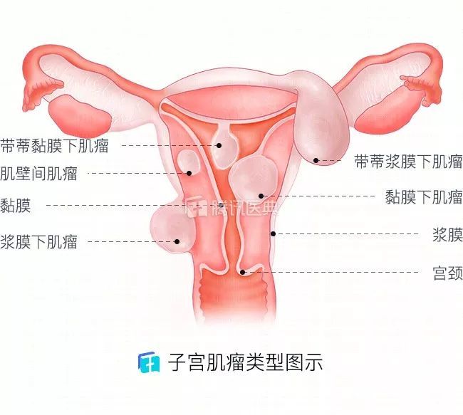 Image source: Tencent Medical Dictionary
 Image source: Tencent Medical Dictionary
Second, estrogen and progesterone – the “motive force” for the growth of fibroids
The exact cause of uterine fibroids is not yet uniform. However, a consensus in the academic community is that estrogen and progesterone are the main promoting factors for the growth of uterine fibroids, and high hormone levels are likely to lead to the occurrence of uterine fibroids.
In reality, uterine fibroids are rare in prepubertal girls. After pregnancy, fibroids will grow with the pregnancy, and after menopause fibroids will gradually shrink.
Studies have also shown that the concentration of estrogen in fibroids is significantly higher than in surrounding normal tissues. This confirms that estrogen and progesterone play an important role in the occurrence and growth of fibroids.
Three, the growth characteristics of fibroids – early silent
Fibroids tend to grow slowly, without any physical response and symptoms in the early stage, and are easily overlooked.
Most women discover fibroids by accident when they do a physical examination over time, and some women discover fibroids after they have grown enough to cause symptoms and go to the hospital for examination.
 Image source: Zhanku Hailuo
Fourth, what does the fibroids look like – the benign surface is smooth and the boundary with the surrounding tissue is clear
Fibroids are nodular “meat balls” raised on the surface of the uterus or in the uterine cavity. They vary in size and are generally smooth and white or light red.
Some fibroids are connected by the pedicle and the uterine wall, and some directly protrude from the smooth uterine surface or the uterine cavity, just like small hills protruding from the flat land.
During an ultrasound or other imaging tests, doctors can tell if these “masses” or “echo areas” are uterine fibroids.
Five, fibroids can degenerate – but there are good and bad points
The incidence of uterine fibroids degeneration is approximately 60%[2]. There are benign and malignant degeneration points, benign are more common, and the probability of malignant transformation (sarcomatoid degeneration) is very low. The occurrence of degeneration has little to do with the location of the fibroids.
 Image source: Zhanku Hailuo
But if it is pregnancy, puerperium, and perimenopause, fibroids are more prone to high incidence. In addition, if the diameter of the fibroids is greater than 5 cm, more prone to degeneration.
Benign degenerations include: hyaline degeneration, cystic degeneration, calcification, and red-like degeneration, mainly due to various reasons resulting in insufficient blood supply to the fibroids themselves, resulting inOccurs when part of the organization loses its original structure. The mechanism of sarcomatoid malignancy remains to be further studied.
6. Can uterine fibroids be prevented?
There is currently no way to absolutely prevent uterine fibroids. However, you can reduce the risk of uterine fibroids by strengthening exercise, eating reasonably, maintaining a good mood, and controlling weight.
 Image source: Zhanku Hailuo
Reviewer: Wang Jianliu| Chief Physician, Department of Obstetrics and Gynecology, Peking University People’s Hospital
References:
[1] Le Jie. Obstetrics and Gynecology [M]. Beijing: People’s Health Publishing House, 2004: 295-298.
[2] Wu Qingmei, Peng Guangxian, Zhou Li. Uterine fibroid degeneration sonogram and its CDFI changes[J]. Journal of Clinical Ultrasound Medicine, 2001, 3(Suppl): 78-80 .
*The content of this article is for the popularization of health knowledge and cannot be used as a specific diagnosis and treatment suggestion, nor can it replace the face-to-face consultation of a licensed physician, and is for reference only.
*The copyright of this article belongs to Tencent Medical Dictionary. Unauthorized reprinting by media is prohibited. Illegal reprinting will be investigated for legal responsibility according to law. Individuals are welcome to forward to the circle of friends.
*Welcome to search on WeChat and pay attention to the public account “Tencent Medical Code” to get more knowledge about health science.