Author: Hu Cheng (The Third Affiliated Hospital of Sun Yat-Sen University)
Image source: Zhanku Hailuo
Common calcium oxalate stones, calcium phosphate stones, and magnesium ammonium phosphate stones, regardless of whether they are infected or not, are generally treated by doctors according to the size of the stones.
1. How to treat when the diameter is less than 5 mm?
If the stone is less than 5 mm in diameter, there is less pain, the color of the urine does not change significantly, and there is little or no infection, the stone will usually pass on its own with the flushing of urine. Such patients must pay attention to life management. When the stone cannot be discharged, the doctor may recommend the use of drugs.
Life Management
Spontaneous removal of stones generally takes 40 days with conservative treatment of life management[1].
Develop good living habits, drink plenty of water, and drink no less than 2500~3000 ml of water per day to promote the excretion of stones with urine.
Drink less coffee, strong tea, carbonated drinks, etc. These drinks contain a lot of caffeine, sugar or artificial additives that can increase the concentration of minerals in the urine and promote the formation of stones.
Simple running, jumping, going up and down stairs, etc., can promote the peristalsis of the ureter, loosen and fall the stones through vibration, which is helpful for stone removal.
Most small ureteral calculi can be excreted by themselves after life management, but it is related to the location of the ureter, and the lower ureteral calculi are easier to excrete. The upper, middle, and lower ureteral excretion rates are 22% [2], 46% [2], and over 90% [1], respectively.
Drug stone removal
If you cannot pass the stones through lifestyle management such as diet and exercise, your doctor may recommend medication. The medicines used vary according to the composition of the stone.
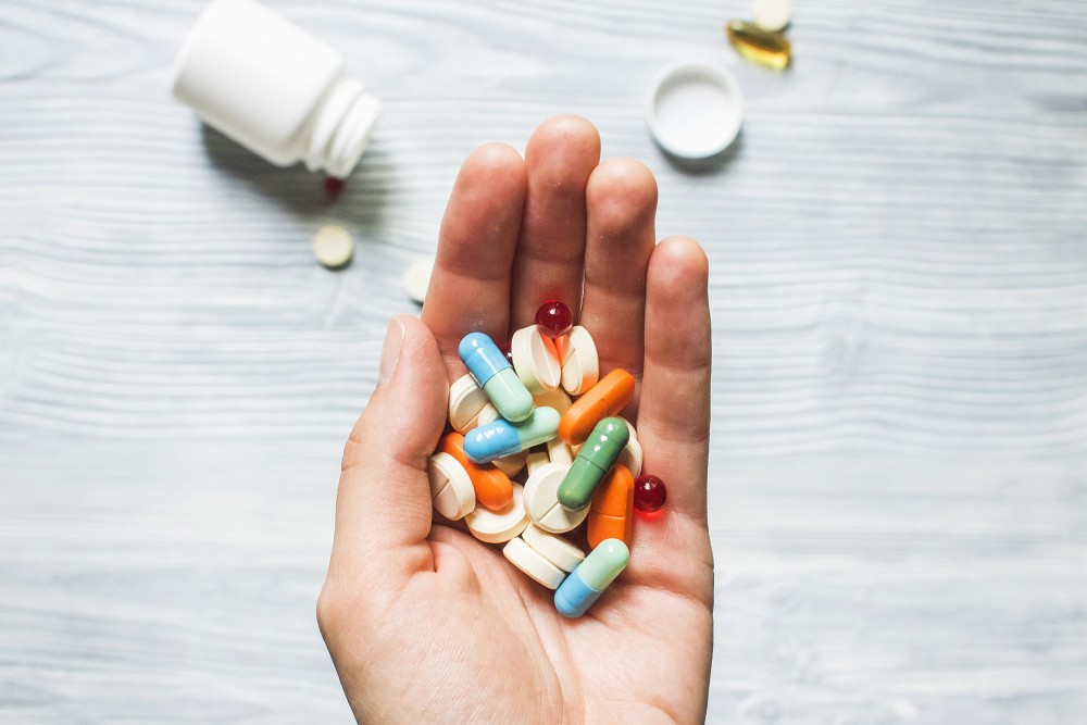
Image source: Zhanku Hailuo
Calcium oxalate stones. Calcium oxalate stones are caused by an increase in calcium in the urine under acidic or neutral conditions. Recommended for patients to use:
Diuretics, such as hydrochlorothiazide, amiloride, etc., can increase the amount of urine and dilute the urine; at the same time, they can also promote the reabsorption of some ions into the blood during the formation of urine, thus reducing the amount of urine Calcium content in the liquid.
Antacids, such as sodium potassium hydrogen citrate, sodium bicarbonate, etc., can make the urine alkaline, which is conducive to the dissolution and discharge of stones.
Calcium phosphate stones
Diuretics may be used, but antacids should not be used because alkaline conditions promote calcium phosphate stone formation.
During the medication process, the pH value of the urine can be detected with a test strip. When the pH value of the urine is greater than 6.5, it is necessary to seek medical attention and adjust the medication plan in time.
Uric acid stones
Antacids, such as sodium potassium hydrogen citrate, sodium bicarbonate, etc., alkalize the urine and promote the dissolution of stones.
Uric acid-lowering drugs, such as allopurinol, febuxostat, etc. During the medication period, it is necessary to go to the hospital regularly to check the blood uric acid level, and increase or decrease the dose in time according to the condition.
Magnesium ammonium phosphate stones. It is a stone caused by bacterial infection, and the following medicines are recommended.
Antibiotics, such as ofloxacin, nitrofurantoin, etc., to treat urinary tract infections.
Urine acidifying drugs, such as ammonium chloride, vitamin C, etc., control the pH of urine below 7.2, which is conducive to the dissolution of stones.
Cystine stones
Antacids, such as sodium potassium hydrogen citrate, sodium bicarbonate, etc., can alkalize urine and promote stone dissolution.
If cystine in urine is too high (greater than 1000mg/L), it is recommended to use penicillamine, tiopronin and other drugs to reduce the concentration of cystine in urine.
2. How to treat stones with a diameter of 5-10 mm?
Stones with a diameter of 5 to 10 mm are relatively large in size, and are easy to get stuck in the ureter, causing pain during large-scale movements, and are not easy to pass out on their own. In this case, in addition to life management, drug expulsion and surgery are generally used in combination [1][2].
Life Management
In addition to drinking more water (no less than 2500~3000 ml per day), drinking less coffee, strong tea, carbonated drinks and other beverages, avoid strenuous exercise when the stone is large to prevent The stone dislodges, scratches or blocks the urinary tract.
Medication
On the basis of life management, drugs are generally used to expel stones. Depending on the composition of the stones, diuretics (such as chlorthalidone, hydrochlorothiazide), antacids ( Such as alkaline citrate, sodium bicarbonate), drugs that reduce uric acid (such as allopurinol, febuxostat), drugs that acidify urine (such as ammonium chloride, vitamin C), etc.
During treatment, pain relievers, anti-infectives, etc. may also be given.
These types of stones are less likely to pass on their own, with only 47 percent passing completely [2]. After the use of drugs, the rate of stone expulsion can be increased to 65% [2].
Surgical stone removal
If there is no obvious effect of drug expulsion, persistent ureteral obstruction, or pain that is difficult to relieve, minimally invasive surgery is required. The doctor will choose the following methods according to the situation.
Extracorporeal shock wave lithotripsy. is the preferred method for the treatment of ureteral calculi. This method is suitable for stones other than cystine, does not require anesthesia, and has fewer complications, but the disadvantage is that there are restrictions on the treatment population, and it is not suitable for pregnant women and people with larger stones, and generally requires multiple treatments. 3]. For upper, middle, and lower ureteral stones, clearance rates were 90%, 84%, and 86%, respectively [1].
Ureteroscopic lithotripsy. Generally used as a second choice after extracorporeal shock wave lithotripsy. Compared with extracorporeal shock wave lithotripsy, the advantage is that it does not require repeated treatment, but the disadvantage is that it causes more short-term postoperative pain, longer hospital stay and more complications than extracorporeal shock wave lithotripsy [4] , the cost is slightly higher.
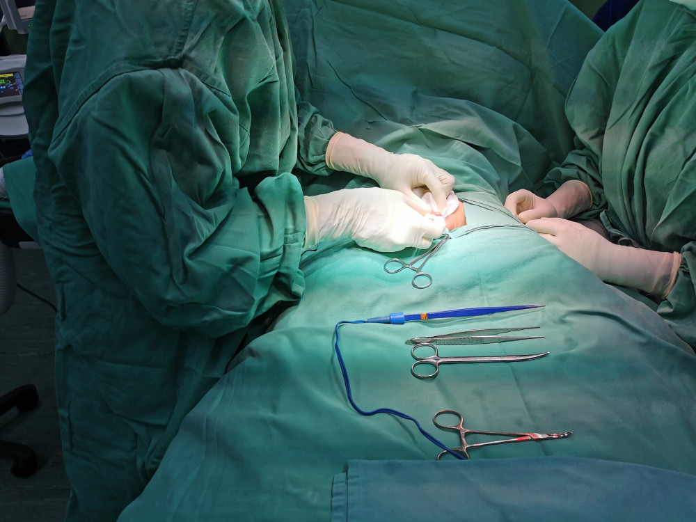
Image source: Zhanku Hailuo
3. How to treat stones larger than 10 mm in diameter?
When the ureteral stone is larger than 10 mm in diameter, it is often easy to get stuck in the ureter, causing urinary tract obstruction and causing hydronephrosis. Continued progression can damage renal function and cause acute renal failure in severe cases. Patients may develop infection, if not controlled in time, it may lead to pyonephrosis, pyomere or even sepsis, which is life-threatening.
For such relatively large stones, drug expulsion has no obvious effect, and surgery is generally required.
Limited water and exercise
Before surgery to relieve urinary tract obstruction, it is not recommended to drink a lot of water, generally no or less water, such as a small sip of water when you are very thirsty. And to take dietary treatment at the same time, avoid drinking strong tea, coffee, etc., or eat foods containing oxalic acid, purines, and high-protein meat. If you drink a lot of water at this time, in the case of increased urine, it cannot be excreted, but it will cause damage to the kidneys. Therefore, those with larger ureteral stones should be treated by surgery in a timely manner as prescribed by a doctor. After the stone is removed, the urine can be successfully excreted, and enough water can be added.
Avoid strenuous exercise to prevent stones from dislodging, scratching or blocking the urinary tract.
When co-infection, surgical drainage + drug anti-infection and pain relief
Pain is usually relieved with pain relievers and antispasmodics. If ureteral stones are complicated by infection, the urine is usually drained first, and the following methods are commonly used.
ureteral stenting. A ureteral stent is a long, thin tube, also known as a “double J tube.” Under the guidance of a cystoscope, the doctor inserts the tube through the urethra, bypasses the stone through the urethra, and extends into the kidney. The other end of the tube remains in the bladder, and urine drains along the stent tube to the bladder. This method is less traumatic to the patient, and the patient can urinate spontaneously.
Percutaneous nephrostomy drainage. This approach is considered when ureteral stenting has failed or is ineffective. A small hole is made in the back, a drainage tube (fistula) is inserted into the kidney, and a bag is attached to the other end to collect urine. Patients do not need to urinate on their own.
Both methods are similar in that they reduce the pressure that urine puts on the kidneys and ureters and relieve pain.
In the case of co-infection, the earlier antibiotics are administered, the better the survival rate[1][4].
Stable stone removal after surgery
Once the infection is under control, the doctor may then perform surgery on the stone.
Extracorporeal shock wave lithotripsy. For stones larger than 10 mm in diameter, the clearance rates were 74%, 76%, and 68% in the upper, middle, and lower ureters, respectively[1].
Ureteroscopic lithotripsy, percutaneous nephrolithotomy. Generally used as a second choice after extracorporeal shock wave lithotripsy. If extracorporeal shock wave lithotripsy is not suitable or the treatment effect is not satisfactory, the doctor will recommend ureteroscopic lithotripsy.
Due to the relatively high cost of percutaneous nephrolithotomy and a certain degree of damage to the kidneys, priority should be given to patients suffering from both kidney stones and urinary tract obstruction at the ureteropelvic junction. Percutaneous nephrolithotomy.
Ureteroscopic lithotripsy for removal of upper, middle, and lower ureteral stones for stones greater than 10 mm in diameterThe clearance rates were 93%, 78%, and 79%, respectively [1], and the nephroscopic clearance rate was 90% to 100% [2]. The complication rates of ureteroscopic lithotripsy and percutaneous nephrolithotomy are 9% to 25% and 15.9%, respectively [4], but the length of hospital stay after nephroscopy is longer [3].
Ureterolithotomy. If the above minimally invasive treatments are ineffective, doctors often choose ureterolithotomy, which can be laparoscopic or “open” surgery. Which type of surgery should be used depends on the specific situation of the patient.
—
References
[1]. Deng Yaoliang. Guidelines for Diagnosis and Treatment of Urological Diseases in China (2014 Edition). People’s Health Publishing House, 2014: 187-208.
[2]. Ordon Michael, Andonian Sero, Blew Brian. et al. CUA Guideline: Management of ureteral calculi. Can Urol Assoc J, 2015, 9: E837-51.
[3]. Zhao Yupei, Chen Xiaoping. Surgery (Volume 1 and 2). Beijing: People’s Health Publishing House. 2016, 10: 752-758, 761-763.
[4]. Wu Mengchao, Wu Zaide, Huang Jiasi. Surgery (Volumes 1, 2 and 2). People’s Health Publishing House, 2008.10: 2338-2344, 2354-2357.
*The content of this article is for the popularization of health knowledge. It cannot be used as a specific diagnosis and treatment recommendation, nor can it replace the face-to-face consultation of a licensed physician. It is for reference only.
*The copyright of this article belongs to Tencent Medical Dictionary. Unauthorized reprinting by media is prohibited. Illegal reprinting will be investigated for legal responsibility according to law. Individuals are welcome to forward to the circle of friends.
*Welcome to search on WeChat and pay attention to the public account “Tencent Medical Code” to get more knowledge about health science.