Experts for this article:
Lv Mingming, Master of Cardiovascular Medicine, North Sichuan Medical College
Hu Houxiang, Chief Physician, Professor, Doctor of Medicine, Master Supervisor, Department of Cardiology, Affiliated Hospital of North Sichuan Medical College
If you were asked to draw a heart
Would your first reaction
draw a ❤️?
But actually
The shape of the heart has nothing to do with the shape of the heart
 < /img>
So
What does the human heart look like?
You may not expect the answer…
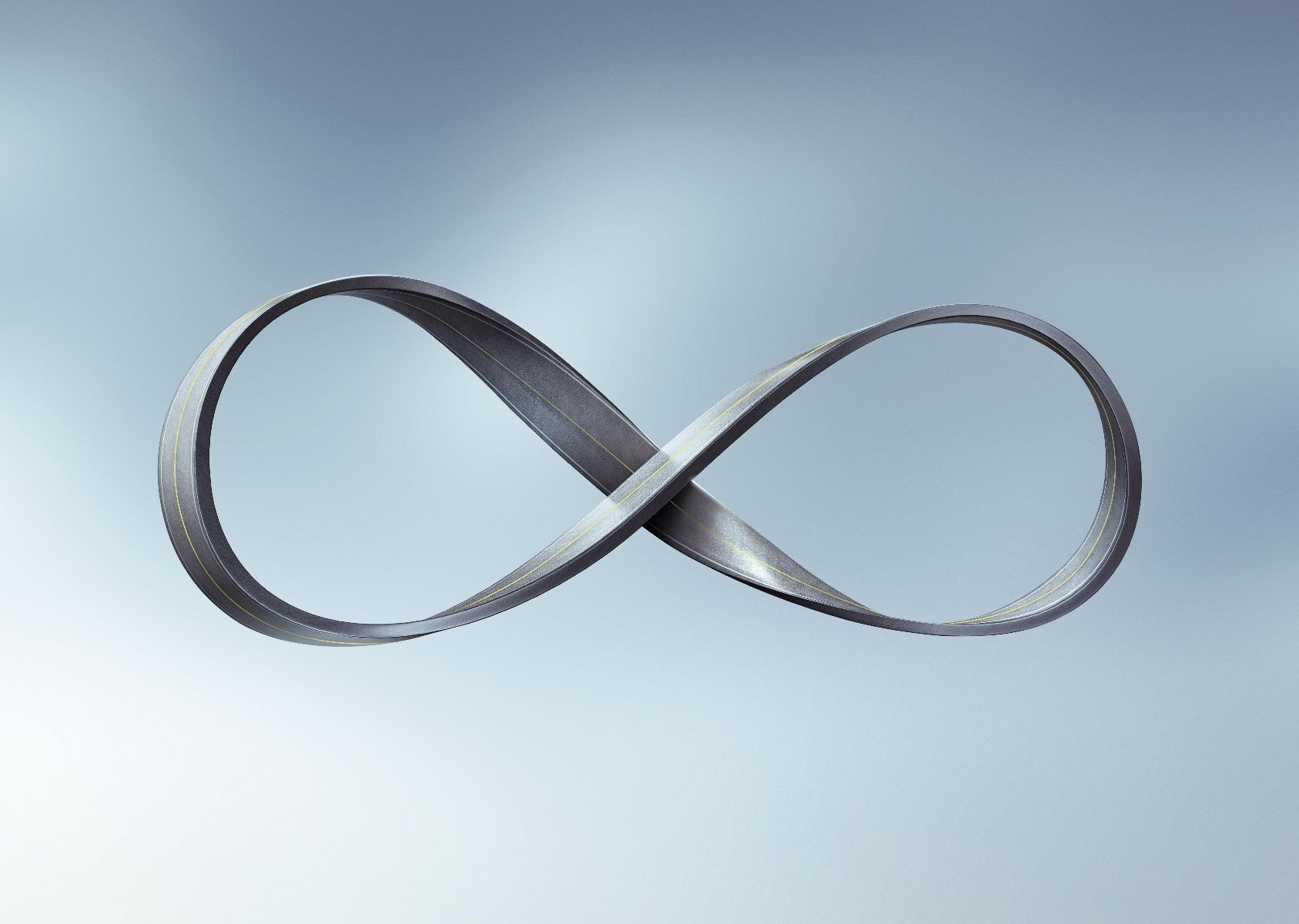
Is the heart a Mobius ring?
Our heart is a hollow organ divided into four chambers, the left and right atrium and ventricle, according to research. But the shape of the heart is not a simple combination of the four chambers. The real shape is a spiral Mobius ring.

As early as the 17th century, some people found that the direction of myocardial fibers was spiral, and subsequent anatomy also found similar findings, but No one knows where this helix begins and ends, let alone the meaning of this helical structure. It wasn’t until half a century ago that Spanish scientist Torrent-Guasp dissected the hearts of nearly a thousand different species to solve this heart anatomy puzzle.
He discovered that the heart is formed by the helical twisting of a broad band of muscle that begins at the root of the pulmonary artery and ends at the root of the aorta. The myocardium is twisted into a figure 8 in a spiral shape, which means that the shape of the heart is similar to a Möbius ring twisted 3 times.
Why is the heart such a complex helical structure?
We all know that structure is the foundation of function.
This helical structure of the heart muscle twists the heart as it contracts, actively pumping blood out and into the heart. This torsional deformation is like a twisting towel-like movement. Systole is the process of twisting and tightening, while diastole is the process of untwisting in the opposite direction after the twisting movement. It can be seen that the heart does not simply contract and relax like a balloon.

So how is such a complex heart formed?
Torrent-Guasp believes that the development of the heart reflects the evolution of the heart from worms to fish, amphibians and finally mammals a billion years ago. For example, around day 18 in a human embryo, a pair of side-by-side heart tubes will form at the head end of the embryo, and then the left and right heart tubes will fuse into a single heart tube, which is the primitive heart, which is similar to the theory that the heart structure is composed of a single muscle band .
The heart tube grows unevenly due to the different growth rates of each segment, and several enlarged areas appear from the head to the tail, which are called cardiosphere, ventricle, atrium and venous sinus in turn. In addition to the growth of the heart tube, there will be a series of changes such as distortion, displacement, and fusion.
At the beginning of week 5, the shape of the heart is almost complete, but the four chambers of the heart have not yet been completely separated. Next, the heart will continue to complete the internal separation of the atrium, ventricle and other structures until the end of the 8th week. If there are obstacles in the separation process, it will cause congenital heart disease, such as atrial septal defect, ventricular septal defect and so on. Therefore, 3-8 weeks of gestation is a critical period for embryonic heart development. During this period, if pregnant women have certain viral infections or exposure to harmful environments, it may lead to the development of fetal heart failure and the formation of congenital heart disease.
How does the heart beat?
Torrent-Guasp distinguishes two longitudinal motions of shortening and elongation, and two transverse motions of constriction and expansion by observing the beating heart, and believes that contraction can only be supported by traveling from top to bottom along the myocardial band. This series of mechanical activities.

Recent studies have found that the electrical signal of normal heart beating originates from the sinoatrial node. The sinus node is located in the right atrium at the bottom of the heart. After the formation of the sinus node, electrical signals are conducted from top to bottom (ie, from the bottom of the heart to the apex), causing the atrium and ventricle to contract and relax successively, that is, the beating of the heart. This also provides further support for Torrent-Guasp’s point.