At present, cerebrovascular disease has become the “number one killer” of Chinese residents, and cerebrovascular disease ranks first in the cause of death in my country, far higher than that in Western countries. In the season of high incidence of stroke, how to understand the situation of cerebrovascular and avoid the occurrence of stroke? There are many methods of cerebrovascular examination, so what is the most authoritative examination method? Next, I will tell you about cerebrovascular examination. “gold standard”.
What is the “gold standard” for diagnosing cerebrovascular disease?
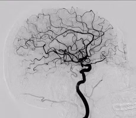
Currently, digital subtraction angiography (DSA) is the “gold standard” for diagnosing cerebrovascular lesions. Whole cerebral angiography is a new clinical examination technique, which can not only clearly display the images of the carotid artery, vertebral artery, large intracranial blood vessels and medium blood vessels of the cerebral hemisphere, but also display the images of the intracerebral veins and capillaries. blood flow, and measuring arterial blood flow. It is one of the most effective ways to check for cerebrovascular disease. This technology has been applied to the examination of cerebrovascular disease, especially for the qualitative location diagnosis of cerebral vascular stenosis, occlusion, aneurysm, arteriovenous malformation, etc., to provide a basis for the prevention and treatment of cerebrovascular disease.
What are the indications for DSA?
①Suspected vascular disease or search for the cause of cerebrovascular disease;
②Suspected cerebral venous disease;
③Cause examination of intracerebral or subarachnoid hemorrhage ;
④ Preoperative examination of blood-rich tumors of head and face;
⑤ To understand the relationship between blood supply and adjacent blood vessels of intracranial space-occupying lesions and the stereotype of some tumors;
⑥ The relationship between vascular lesions and surrounding anatomy should be clarified before vascular intervention or surgical treatment;
⑦ Arterial thrombolysis or other intravascular treatment is required for acute cerebrovascular disease;
⑧ head Post-treatment review of facial and intracranial vascular disease.
What are the contraindications to DSA?
①Allergy or intolerance to iodine contrast agent;
②Allergy to interventional equipment;
③Severe heart, liver, and renal insufficiency;
④Local infection of the puncture point;
⑤Complication of brain herniation. Special circumstances can be discussed by all parties, and informed consent can be individualized.
How does DSA work?
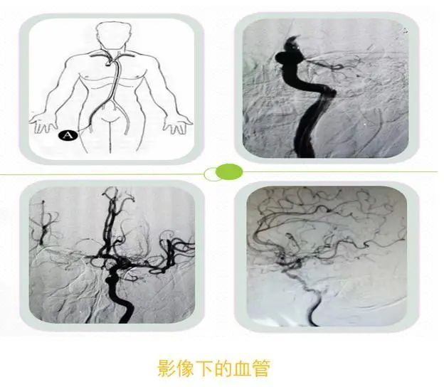
By injecting the iodine-containing contrast agent into the internal carotid artery, vertebral artery or femoral artery, the different time, travel and distribution of the contrast agent entering the brain along with the blood circulation are recorded by continuous X-ray film. Visualize blood vessels.
DSA has so far been regarded as the gold standard for cerebrovascular imaging, and there is currently no test for cerebrovascular disease. The diagnostic accuracy is better than DSA.
In general: through DSA examination, we can accurately understand the number, location, size, shape of vascular lesions, and the relationship with surrounding blood vessels; also can preliminarily predict or understand the development of diseases, such as bleeding risk of infarction, risk of infarction, etc.