“I have a heart attack!”
“Hurry up and have a cardiac imaging! Cardiac imaging is the most accurate, and it can detect heart disease in one fell swoop!”
Do you often hear such conversations, and do many people think so too: Cardiac angiography is the most accurate heart examination!

Of course not!
Cardiac imaging is not the most accurate test for heart disease, because the heart is divided into several parts, and different parts are examined differently:
One, the composition of the heart

1. The water system, which is the blood vessels of the heart.
2. The circuit system, which is the beating of the heart.
3. The structure of the heart, that is, the valves, myocardium, size, etc. of the heart.
Second, different departments have problems, and the inspection methods are different
1. Is there plaque, stenosis, or blockage in the blood vessels of the heart?
What tests should be done when there is suspected plaque or narrowing, or blockage in a blood vessel in the heart?
1), cardiovascular plaque
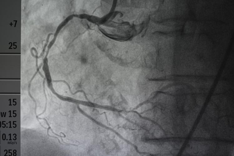
When there is suspected plaque in the cardiovascular system, the commonly used examination is coronary CT, also called coronary CTA or enhanced cardiac CT, or the familiar cardiac angiography.
But both coronary CT and coronary angiography have radiation and contrast agents, so they are not routine examinations. In particular, coronary angiography is still an invasive examination, and the risk is relatively greater, so a coronary CT or coronary angiography is only recommended when necessary to check whether there is plaque or stenosis in the heart blood vessels.
So cardiography is an accurate way to check for stenosis of blood vessels in the heart, not an accurate way to check the heart. Because the heart is about the circuit system of the heart or the overall structure of the heart, it is impossible to examine the heart by angiography alone.
2), heart blockage
Heart blockage, generally refers to complete blockage of the heart, that is, acute myocardial infarction, which is generally diagnosed by two of the three items of the patient’s persistent angina pectoris symptoms + ECG changes + myocardial enzymes.
If myocardial infarction is highly suspected or diagnosed, coronary angiography can be performed, on the one hand, to further identify the location of blood vessel blockage, on the other hand, to open blood vessels, and angiographic intervention methods are also required.
2. The heartbeat problem
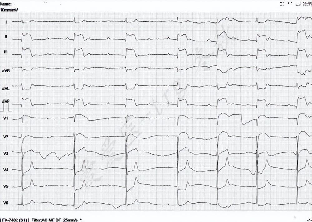
Whether the heartbeat is too fast, the heartbeat is too slow, or the heartbeat is too chaotic. Generally, there is a problem with the cardiac circuit system. At this time, an electrocardiogram is the first choice.
But many times, when we arrive at the hospital, our heartbeat is not so fast, not so slow, and not so chaotic. Therefore, it is difficult to find problems through ordinary ECG examination, so at this time, a dynamic ECG, which is a 24-hour ECG, is required.
If a 24-hour ECG does not reveal a problem, and arrhythmia is still highly suspected, then electrophysiological testing may be considered.
For example, the premature beats, atrial fibrillation, atrial tachycardia, supraventricular tachycardia, atrioventricular block, ventricular tachycardia, etc. that we often say can only be found by ECG examination.
Of course, the electrocardiogram, as we said above, can also play a huge and even irreplaceable role in acute myocardial infarction.
3. Heart structure
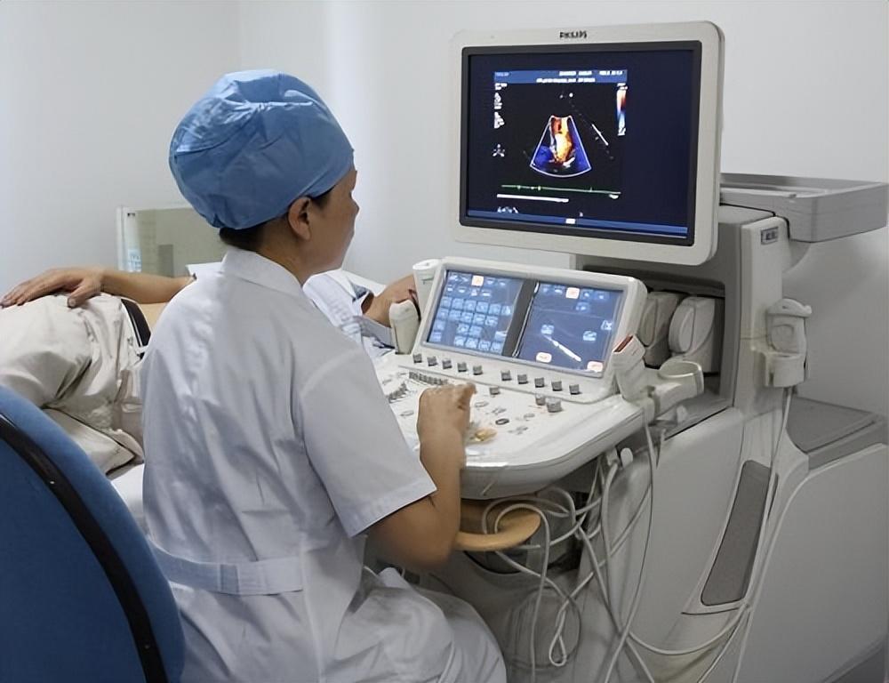
We often say that the heart is a house, the blood vessels are the pipes, the beats are the circuits, and the mechanism of the heart is the room, the walls, and the functions.
This part of the heart problem depends on echocardiography, which can find out whether there are problems with the mitral valve, tricuspid valve, aortic valve, pulmonary valve, etc. of the heart; the left side of the heart Whether the size of the atrium, right atrium, left ventricle, or any ventricle is normalized; whether the myocardium is thickened or thinned; whether the ejection function of the heart is normal, whether there is heart failure, etc., are generally checked by cardiac color Doppler ultrasound.
4. Heart blood test

You may have heard of myocardial enzymes, thinking that if the myocardial enzymes are normal, there will be no myocardial infarction, and if the myocardial enzymes are abnormal, myocardial infarction will occur.
Of course not.
When a myocardial infarction occurs, cardiac enzymes are elevated only for a certain period of time. In other words, myocardial enzymes were normal an hour before myocardial infarction, and normal after a few days. Therefore, normal myocardial enzymes does not mean that there is no myocardial infarction.
Furthermore, even if myocardial enzymes are high, it does not mean myocardial infarction, because myocarditis, myocardial damage and other problems will cause myocardial enzymes to rise.
Therefore, myocardial enzymes must also be comprehensively judged in combination with the specific situation of the patient.
In short, cardiac angiography is not the most accurate way to check for heart disease, but a more commonly used and more accurate method to check whether there is stenosis in the blood vessels of the heart.
Which tests are needed for the specific heart must be determined after comprehensive consideration based on the specific symptoms of the patient.
The general principle is: do not open the inspection!